Page created on October 3, 2018. Last updated on January 20, 2019 at 11:23
Organ: Heart
Description: We can see three slices of the heart. The ventricular wall of the left ventricle is more than 15 mm, meaning it is increased compared to normal. The lumen is very small as well.
Diagnosis: Concentric hypertrophy of the left ventricle
Causes:
- Increased afterload
- Systemic hypertension
- Aortic stenosis
Theory:
This topic is best understood in tandem with preparation 18.
When there is increased afterload the left ventricle must push out the blood into the aorta against a stronger pressure than if the afterload was normal. The ventricle responds to the increased afterload by concentric hypertrophy. In this type of hypertrophy new sarcomeres are added parallelly to the long axis of the myocytes, so that the muscle fibre increases in diameter. This causes the ventricular wall to increase in thickness. This increase in muscle mass allows the ventricle to contract with more force.
The muscle fibres will continue to grow to keep up with the afterload. While the muscle fibres increase in diameter, the vessels do not. The vessels therefore can’t supply the continuously enlarging muscle fibres. The fibres will eventually stop growing as they experience hypoxia because of hypoperfusion.
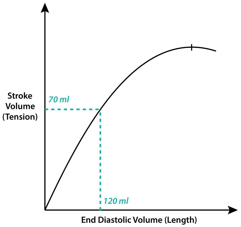
Because the ventricle no longer can increase its muscle size to keep up with the afterload, it must change strategies. Recall that the Frank-Starling mechanism of the heart causes the heart to increase its contractility in response to an increased end-diastolic volume. The ventricle will then start to undergo dilatative hypertrophy to dilate the lumen to increase the EDV. However, this dilation causes a lot of strain on the ventricle, as we will see.
The Laplace law states that wall tension is proportional to the pressure inside the ventricle times the radius of the ventricle, like this:
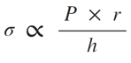
According to this law, when the radius of the ventricle increases, the tension in the ventricular wall must increase to maintain the same pressure. In simpler words, when the ventricle dilates enough the increasing wall tension will make it harder for the ventricle to contract, which will decrease the contractility. This is what causes the “drop-off” on the end of the Frank-Starling curve.
The ventricle has now run out of strategies (compensation mechanisms) to keep up the contractility, so it will eventually to pump out enough blood, causing the heart to become decompensated.
So, to sum up:
- Increased afterload causes concentric hypertrophy
- The muscle fibres increase in size, but the vessels can’t, so the muscle fibres eventually stop growing because of a lack in blood supply
- Because the muscle fibres can’t keep growing the ventricle must compensate for the increased afterload somehow else.
- To take advantage of the Frank-Starling mechanism the ventricle will start to dilate by dilatative hypertrophy.
- The Laplace law states that increased dilation of the ventricle causes increased strain on the ventricular wall. This increased strain causes a decrease in contractility.
- The decreased contractility eventually results in the ventricle not being able to compensate for failing heart, causing decompensation.
Please check the Laplace law you’ve written about. h is the width of the ventricule. Also I really don’t know what sign after sigma is there. It schould be only a =. Keep up the good work!
∝ means proportionality. It means that σ isn’t directly equal to the right side of the equation, but it’s proportional to it.
I did add the note about h though. Thanks!