Table of Contents
Page created on May 22, 2019. Last updated on December 18, 2024 at 16:56
I recommend reading through the internal medicine topic 3 for a better introduction to this topic.
Introduction
Blocks of the conduction system doesn’t happen only at the SA or AV nodes; it can happen in the conduction system inside the ventricles as well. These are called intraventricular blocks and include:
- Right bundle branch block (RBBB)
- Left bundle branch block (LBBB)
- Left anterior fascicular block (LAFB) or left anterior hemiblock
- Left posterior fascicular block (LPFB) or left posterior hemiblock
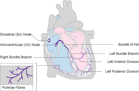
As the names imply, RBBB is a block of the right bundle branch and LBBB is a block of the left bundle branch. The left bundle branch is divided into two branches, the anterior fascicle and the posterior fascicle. LAFB and LPFB involves block of these fascicles, respectively.

The word “division” is used instead of “fascicle”. From https://www.nottingham.ac.uk/nursing/practice/resources/cardiology/function/conduction.php
Some of these blocks can be rate-dependent, meaning that they only appear during high heart rates. This is more common in RBBB than in the other types.
These conditions are not as severe in themselves, but they often indicate that there has been local ischaemia or degeneration at the site of the block. Cardiac output may be decreased due to loss of ventricular synchronicity.
Right bundle branch block
When the right bundle branch is blocked the conduction signal must travel to the right ventricle through the myocardium rather than through the right bundle branch. This is much slower and so the right ventricle is depolarized later than the left ventricle. This causes the QRS complex to become wider than normal.
Etiology:
- Atrial septal defect
- Right ventricular hypertrophy
- Right ventricular volume load
- Pulmonary embolism
- Ischaemic heart disease
- Myocardial infarct
- Myocarditis
The right bundle branch is vulnerable and easily damaged. RBBB can also appear in healthy persons.
ECG morphology:
Some of these signs are only seen in V1 and V2, the leads that are closest to the right ventricle, where the defect lies.
- Widened QRS (> 120 ms)
- VAT (ventricular activation time) > 40 ms
- rSR’ formation (M-shaped or “rabbit-ear” shaped QRS) in V1, V2
- Wide S wave in lead I, II, aVL
- Secondary repolarization disorder
Left bundle branch block
When the left bundle branch is blocked the conduction signal must travel to the left ventricle through the myocardium rather than through the left bundle branch. This block occurs in the part of the left bundle branch proximal to where it divides into the anterior and posterior fascicles.
Etiology:
- Ischaemic heart disease
- Myocardial infarct
- Left ventricular hypertrophy
LBBB never occurs in healthy hearts, as the left bundle branch is not easily damaged.
ECG morphology:
Some of these signs are only seen in V5 and V6, the leads that are closest to the left ventricle, where the defect lies.
- Widened QRS (> 120 ms)
- VAT > 40 ms
- QS or rS complex in V1, V2
- Lack of Q-wave in V5, V6
- Wide R-wave in V5, V6
- rSR’ formation (M-shaped or “rabbit-ear” shaped QRS) in V5, V6
- Secondary repolarization disorder
Left anterior hemiblock
Left anterior hemiblock occurs when the anterior fascicle of the left bundle branch is blocked. This causes the posterobasal part of the left ventricle to be depolarized before the anterior part.
Patients are usually asymptomatic and require no treatment. If there is no underlying organic heart disease isolated left anterior hemiblock is considered benign.
ECG morphology:
- Extreme left axis deviation
- Normal QRS
- qR complex in lead I
- rS complex in lead III
Left posterior hemiblock
Left posterior hemiblock occurs when the posterior fascicle of the left bundle branch is blocked. This causes the anterior part of the left ventricle to be depolarized before the posterobasal part. This is much less frequent than left anterior hemiblock.
Patients are usually asymptomatic and require no treatment. If there is no underlying organic heart disease isolated left posterior hemiblock is considered benign.
ECG morphology:
- Extreme right axis deviation
- Normal QRS
- rS complex in lead I
- qR complex in lead III
Lenegre syndrome
According to the book, Lenegre syndrome refers to the presence of both RBBB and left anterior hemiblock. It explains this common presence by the fact that these two conduction systems share a blood supply.
If you try to google Lenegre syndrome you will not find any international sources that agree with the book. International sources call it a genetic defect in a sodium channel, leading to fibrosis of the conducting network, and not RBBB and left anterior hemiblock specifically.
If you google Lenegre syndrome in Hungarian, you will find some Hungarian sources that agree with POTE’s definition, mostly other universities. Hungarians call it “jobb Tawara szár blokk” (JTSZB) and bal anterior hemiblock (BAH).