Page created on April 24, 2018. Last updated on December 18, 2024 at 16:55
Immunohistochemistry

Immunohistochemistry uses the antigen-antibody interaction to detect a specific antigen in a tissue. We add an antibody that has an enzyme attached to it to the tissue. We also add the substrate for this enzyme and an inactive color molecule called the chromogen. When the antibody binds its antigen in the tissue, the enzyme will convert the enzyme substrate into a different product. This product will then activate the color product, making it colorful.
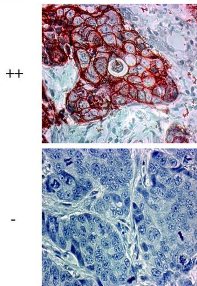
This colorful reaction can be seen in the microscope and can be used to tell whether a certain type of cancer has a specific antigen or not, which can be important for the treatment. It results in images like these.
Both slides are of a breast cancer. The upper cancer is positive for an antigen called HER2, while the lower cancer is negative for the same antigen. The treatments will therefore be different.
Immunofluorescence
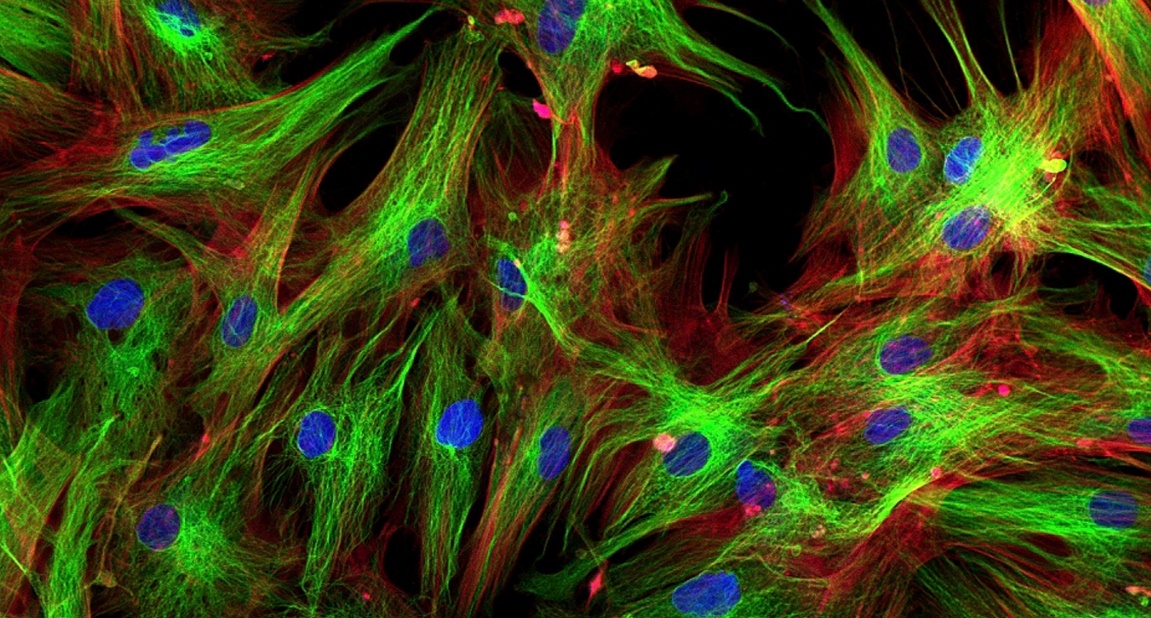
In this technique antibodies are also used and are also attached with something. In this case however, the antibodies are attached to a type of molecule called fluorochrome and not an enzyme. A fluorochrome is a molecule that emits a specific color when a specific wavelength of light is shined upon it, which is done by a special microscope called a fluorescent microscope. It results in images like this (after photoshop):
