Table of Contents
Page created on July 14, 2020. Last updated on July 22, 2020 at 21:28
Learning objectives
- What is the origin of the female gamete?
- What is the origin of the primary oocyte, and what is its DNA content?
- What is the origin of the secondary oocyte, and what is its DNA content?
- What is the origin of the mature ovum, and what is its DNA content?
- Describe the primordial follicle
- Describe the primary follicle
- Describe the antral follicle
- Describe the graafian follicle
- What is the zona pellucida, and what is its function?
- What is the cumulus oophorus, and what is its function?
- What is the function of the theca interna cells?
- What is the function of the granulosa cells?
- What marks the beginning of the menstrual cycle?
- What occurs during the follicular phase of the ovarian cycle?
- What occurs during the desquamation phase of the endometrial cycle?
- What occurs during the proliferation phase of the endometrial cycle?
- When does ovulation occur?
- What occurs during the luteal phase of the ovarian cycle?
- What occurs during the secretory phase of the endometrial cycle?
Oogenesis
Oogenesis refers to the development of an immature oogonium into a secondary oocyte which is ready to be fertilized. Oogenesis begins during the embryonic development of the female. This means that a pregnant woman who carries a girl carries not only the next generation as a foetus, but the next generation after that as eggs.
Chromosome count
The most complicated and difficult concept to understand is how the amount of DNA changes with each round of meiosis I and meiosis II, especially considering that these processes are interrupted and paused for many years during oogenesis.
Humans are diploid organisms, meaning that we have two copies of each chromosome in each cell, one from the father and one from the mother. This includes 23 chromosomes from each parent, for a total of 46 chromosomes. The number of copies of chromosomes in each cell is denoted by the letter n. Human cells are 2n, except for gametes.
Before a cell divides the DNA in the cell is replicated. The amount of DNA in the cell doubles, but the number of chromosomes remain the same, but the chromosomes have gotten bigger – instead of being “lines” they are now X-shaped, i.e. two lines joined at the centre. The X-shaped chromosomes are comprised of two sister chromatids, which are identical.
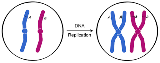
The amount of DNA before and after DNA replication.
Another letter, c, denotes the amount of DNA in the cell. After replication the number of chromosomes is the same, but the amount of DNA has doubled, as two sister chromatids have been formed. Before replication the cell is 2n2c, but after replication (and before cell division) the cell is 2n4c. After cell division the sister chromatids are separated, forming two 2n2c cells.
Meiosis
Meiosis is the process where one 2n2c mother cell replicates its DNA to become 2n4c, after which it divides into four 1n1c cells. Meiosis is divided into two processes, meiosis I and meiosis II.
During meiosis I the DNA of the 2n2c mother cell is first replicated so that it becomes 2n4c. During the prophase, metaphase, and anaphase of meiosis I the cell remains 2n4c. During telophase the cell divides, forming two 1n2c daughter cells.
During meiosis II these 1n2c cells remain 1n2c until the telophase, at which point they divide to form four 1n1c daughter cells, and the process of meiosis is complete.
Polar bodies
It’s important to note that, of the four daughter cells, only one develops into a mature gamete. This is because only one of the daughter cells receives most of the cytoplasm of the mother cell; the remaining three receive barely any cytoplasm. They are called polar bodies. They’ll degenerate.
Oogenesis in the foetal period
During the foetal period of the female so-called primordial germ cells migrate to her primordial gonads. Here they differentiate into oogonia. At the 4th week of gestation these oogonia begin to replicate by mitosis. From the 4th week of gestation until month 7 of gestation these oogonia continue to replicate.
During this period some oogonia start the process of meiosis I, at which point they’re known as primary oocytes. These primary oocytes don’t complete meiosis I but are arrested in the prophase I of meiosis I. After month 7 all oogonia have either matured into primary oocytes or died.
Oogenesis after the foetal period
The primary oocytes remain arrested in prophase I up until the time when ovulation nears. A surge in LH approximately one day before ovulation causes the primary oocyte to finish meiosis I, at which point it becomes a secondary oocyte.
The secondary oocyte continues with meiosis II but is arrested in the metaphase approximately 3 hours before ovulation. 3 hours later the secondary oocyte is ovulated.
If the secondary oocyte is fertilized it will complete meiosis II, yielding the mature ovum. If it is never fertilized the cell will simply degenerate, never having finished meiosis II.
The table below shows the period, events, cell type, and DNA content of the oocytes as they mature.
|
Period |
Events | Cell | DNA content |
| 4th week of gestation | Oogonia replicate by mitosis | Oogonia |
2N2C |
|
From 4th week of gestation to 4 weeks before birth |
Primary oocytes start meiosis I but are arrested in prophase | Primary oocytes | 2N4C |
| Right before ovulation | Meiosis I resumes and finishes, forming secondary oocytes. They initiate meiosis II but are arrested in the metaphase | Secondary oocytes |
1N2C |
|
After fertilization |
The secondary oocyte finished meiosis II, forming the mature ovum | Mature ovum |
1N1C |
Folliculogenesis
The oocyte lives inside an ovarian follicle its whole life. Each follicle contains a single oocyte. As the oocyte matures and develops from an oogonium to an ovum the follicle matures with it, progressing from a primordial follicle until a graafian follicle.
Primordial follicle
During oogenesis in the foetal period, while the oogonia are replicating by mitosis, each oogonium is surrounded by flat epithelial cells which originate from the surface epithelium of the ovary. These epithelial cells are follicular cells and will give rise to the ovarian follicle.
The primary oocyte together with its surrounding follicular cells is known as a primordial follicle. It is the follicular cells which arrest the primary oocyte in the prophase of meiosis I. At month 7 there are only primordial follicles with primary oocytes in the ovary. No oogonia remain.
Primary follicle
As the primordial follicle grows the flat follicular cells differentiate into cuboidal granulosa cells. The granulosa cells rest on a basement membrane which separates them from the surrounding ovarian stroma. Cells from the ovarian stroma will form the thecal layer (theca folliculi) on the outside of the basement membrane. The cells of the theca folliculi organize into an inner layer of cells called the theca interna cells, and an outer fibrous capsule, the theca externa.
The granulosa cells and the oocyte secrete glycoproteins onto the surface of the oocyte, forming the zona pellucida, a layer of glycoproteins around the oocyte.
At puberty primordial follicles continuously mature into primordial follicles.
Antral follicle
Also called the vesicular follicle, the antral follicle is characterised by the formation of an antrum, a cavity, in the follicle. As follicular development continues fluid-filled spaces appear between the granulosa cells. These spaces coalesce to form this antrum. The formation of the antrum pushes the oocyte off centre.
The granulosa cells which surround the oocyte form a structure called the cumulus oophorus.
Every menstrual cycle 15 – 20 primary follicles begin to mature. Only some reach the antral follicle stage; the others die.
Graafian follicle
Also called the mature vesicular follicle, the graafian follicle is the most mature stage. As the antrum continues to grow the follicle grows in size; the graafian follicle is 25 mm in diameter, quite visible to the naked eye.
Approximately 37 hours before ovulation the antral follicle has matured into the graafian follicle.
The table below shows the stages of follicles and their description.
|
Period |
Stage of follicle | Stage of oocyte | Description of follicle |
| Month 7 of gestation – puberty | Primordial follicle | Primary oocyte |
Primary oocyte surrounded by flat epithelial follicular cells |
|
Puberty – start of ovarian cycle |
Primary follicle | Primary oocyte | Primary oocyte surrounded by zona pellucida, cuboid granulosa cells, the theca interna, and the theca externa |
| During the follicular phase of the ovarian cycle | Antral follicle | Primary oocyte |
An antrum has formed between granulosa cells. The granulosa cells around the primary oocyte form the cumulus oophorus. |
|
37 hours before ovulation – ovulation |
Graafian follicle | Primary oocyte
Secondary oocyte (3 hours before ovulation) |
The follicle is 25 mm, as the antrum has grown. |
The menstrual cycle
At puberty females begin to experience their menstrual cycle. Their first menstrual cycle is called the menarche, which occurs at around 12 – 13 years. This cycle affects both the ovaries and the endometrium of the uterus.
During each cycle one mature oocyte is released from the ovaries, from which it will travel through the fallopian tubes and eventually into the uterus. If the mature oocyte was fertilized by a sperm cell the blastocyst will implant into the endometrium. If not fertilized the oocyte will be shed.
Each menstrual cycle lasts approximately 28 days, with anything between 21 and 35 days being normal. The cycle starts the day of the menstrual bleeding, which is denoted day 1.
As stated earlier the menstrual cycle affects both the ovaries and the endometrium. Thus, the menstrual cycle is comprised of two cycles which occur simultaneously: the ovarian cycle and the endometrial or uterine cycle.
Control of the menstrual cycle
The menstrual cycle is controlled by hormones. The secretion of these hormones is under the control of two very important parts of the brain, the hypothalamus and the anterior lobe of the pituitary gland (also called the adenohypophysis). In general, the hypothalamus instructs the anterior pituitary which hormones to produce and when, and the anterior pituitary synthesises and releases these hormones into the blood. The hormones then reach the ovaries. Because the hypothalamus controls the anterior pituitary which controls the ovaries, we call this cooperation of organs the hypothalamic-pituitary-gonadal axis.
This axis is based on the following concept: The hypothalamus produces a hormone called gonadotropin-releasing hormone (GnRH). Like its name implies this hormone stimulates the release of gonadotropins from the anterior pituitary.
There are two gonadotropins, follicle-stimulating hormone (FSH) and luteinizing hormone (LH). These hormones have different functions.
Follicle-stimulating hormone
FSH, like the name implies, stimulates the maturation of the ovarian follicles. These maturing follicles will produce oestrogens, the female sex hormone, by the following mechanism:
The theca interna cells of the follicle produce androgens like testosterone and androstenedione. These male sex hormones will then travel to the granulosa cells which converts them androstenedione and testosterone into female sex hormones (oestrogens) like oestrone and oestradiol respectively, with the help of the enzyme aromatase.
Actually, the production of androgens by the theca cells is not stimulated by FSH, but the conversion of the androgens into oestrogens by the granulosa cells is.
Luteinizing hormone
The role of LH is less self-explanatory than that of FSH. Right before day 14 (midcycle) there is a surge in LH release from the anterior pituitary. This has multiple effects:
- LH causes the oocytes to complete meiosis I and initiate meiosis II.
- LH stimulates the production of progesterone by follicular stromal cells.
- LH stimulates collagenase, an enzyme which digests collagen fibres surrounding the follicle
- LH increases local prostaglandin levels in the ovary, causing local muscle contractions in the ovarian wall. This extrudes the oocyte of the graafian follicle. This is the ovulation.
Days 1 – 14
The follicular phase of the ovarian cycle
The follicular phase of the ovarian cycle takes place in the first half of the menstrual cycle. FSH stimulates the development of several follicles of the ovaries. Approximately 15 – 20 primary follicles will mature into antral follicles, but only one will mature into a graafian follicle.
The other follicles will degenerate and become atretic. The atretic follicles and their oocytes will degenerate and be replaced by connective tissue, forming a corpus atreticum.
The desquamation (or menstrual) phase of the endometrial cycle
Because no pregnancy occurred during the last cycle, the corpus luteum degenerates, and along with it the progesterone levels decrease. This induces vasospasms in the uterine spiral arteries, which causes the compact and spongy layers of the endometrium to die due to being deprived of blood (ischaemia).
This occurs during the first four days of the endometrial cycle. The dead layers of the endometrium leave the body through the vagina along with blood, causing the menstrual bleeding. Only the basal layer of the endometrium remains.
The proliferation phase of the endometrial cycle
The oestrogens produced by the ovarian follicles stimulates proliferation of the basal layer of the endometrium. This proliferation eventually forms new spongy and compact layers.
Ovulation (day 14)
Ovulation usually occurs in the middle of the cycle, at day 14. In the days leading up to day 14 the ovarian follicle has reached the graafian stage. A surge in LH approximately one day before ovulation stimulates multiple processes which altogether cause the secondary oocyte to be extruded from the graafian follicle, which is the ovulation.
LH stimulates collagenase, an enzyme which digests collagen fibres surrounding the follicle. LH also increases the level of prostaglandins in the ovary, which causes local muscle contractions in the ovarian wall. At day 14 these processes cause the secondary oocyte to be extruded from the graafian follicle together with the cumulus oophorus.
The granulosa cells comprising the cumulus oophorus form the corona radiata around the zona pellucida. The corona radiata functions as a barrier which spermatozoa must penetrate.
Days 15 – 28
The luteal phase of the ovarian cycle
After ovulation the remains of the graafian follicle develops into the corpus luteum under the influence of LH. The corpus luteum produces progesterone, a hormone which aids pregnancy.
If pregnancy occurs the syncytiotrophoblast of the embryo will produce hCG, a hormone which keeps the corpus luteum alive, allowing it to keep producing progesterone. In this case the corpus luteum will grow into a corpus luteum of pregnancy.
If no pregnancy occurs the corpus luteum will eventually degenerate due to lack of stimulation from hCG. It degenerates into a mass of fibrotic scar tissue called the corpus albicans.
The secretory phase of the endometrial cycle
The progesterone produced by the corpus luteum causes the new functional layer of the endometrium to change in preparation of implantation by the fertilized egg. The uterine spiral arteries grow deeper, and the endometrium start to produce glycogen-rich secretions. This provides a good environment for the egg to implant if fertilization occurs.
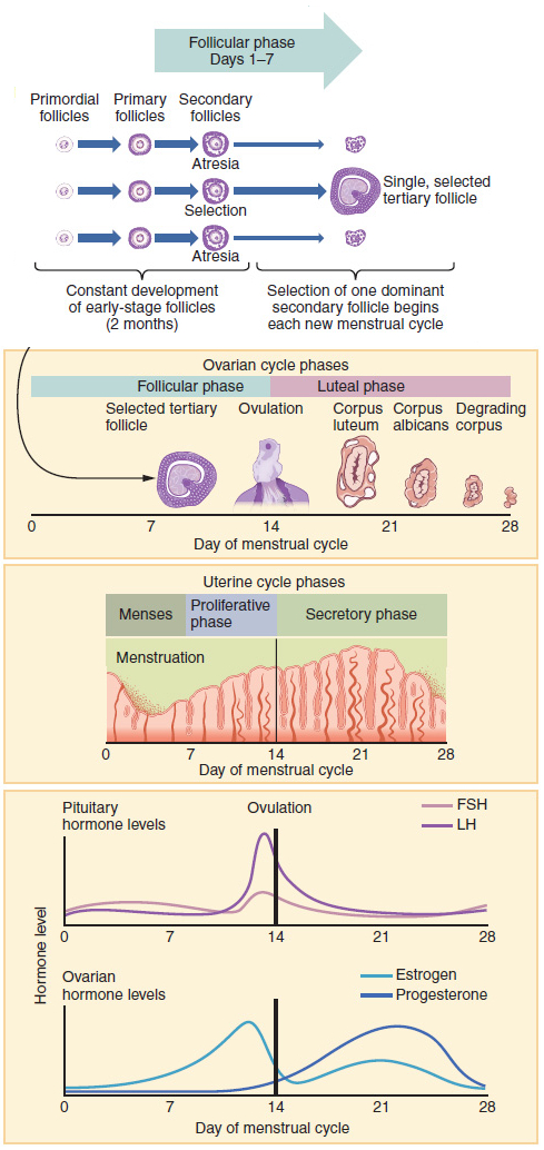
A graphical representation of the menstrual cycle. From Wikipedia.
Summary
- What is the origin of the female gamete?
- From primordial germ cells which migrate to the primordial gonads
- What is the origin of the primary oocyte, and what is its DNA content?
- The primary oocytes is an oogonium which has started meiosis I but is arrested in the prophase
- After month 7 of gestation all oogonia have become primary oocytes
- 2N4C
- What is the origin of the secondary oocyte, and what is its DNA content?
- The secondary oocyte is a primary oocyte which has started meiosis II but is arrested in the metaphase
- The maturation from primary to secondary oocyte is stimulated by the LH surge at day ~13
- 1N2C
- What is the origin of the mature ovum, and what is its DNA content?
- The mature ovum is a secondary oocyte which has finished meiosis II after being fertilized by a sperm cell
- 1N1C
- Describe the primordial follicle
- The primordial follicle consists of a primary oocyte surrounded by epithelial follicular cells
- Describe the primary follicle
- The primary follicle consists of a primary oocyte surrounded by cuboidal granulosa cells, a basement membrane, theca interna, and theca externa cells
- During puberty all primordial follicles have matured into primary follicles
- Describe the antral follicle
- The antral follicle is a primary follicle with an antrum between the granulosa cells
- The granulosa cells around the primary oocyte form the cumulus oophorus
- Some primary follicles mature into antral follicles during the follicular phase of the ovarian cycle
- Describe the graafian follicle
- The graafian follicle is an antral follicle in which the antrum has grown, making the follicle 25 mm in diameter
- Only one antral follicle matures into a graafian follicle each ovarian cycle
- What is the zona pellucida, and what is its function?
- It is a layer of glycoproteins around the oocyte
- It initiates the acrosome reaction
- What is the cumulus oophorus, and what is its function?
- The cumulus oophorus is a layer of granulosa cells which surround the oocyte in the antral and graafian follicle
- The cumulus oophorus becomes the corona radiata after ovulation
- What is the function of the theca interna cells?
- Theca interna cells produce androgens like androstenedione and testosterone
- What is the function of the granulosa cells?
- Granulosa cells convert androstenedione and testosterone into oestrone and oestradiol with the help of aromatase
- What marks the beginning of the menstrual cycle?
- The first menstrual bleeding of the cycle
- What occurs during the follicular phase of the ovarian cycle?
- Day 1 – 14
- FSH is released from the anterior pituitary, causing 15 – 20 primary follicles to mature
- Only one of them will reach the graafian follicle stage; the rest become atretic
- What occurs during the desquamation phase of the endometrial cycle?
- Day 1 – 4
- Due to the lack of pregnancy the corpus luteum degenerates, causing the compact and spongy layers of the endometrium to shed
- What occurs during the proliferation phase of the endometrial cycle?
- Day 5 – 14
- The oestrogens produced by the ovarian follicles stimulates proliferation of the basal layer of the endometrium
- When does ovulation occur, and what event triggers it?
- Day 14
- Ovulation is triggered by the LH surge
- What occurs during the luteal phase of the ovarian cycle?
- Days 15 – 28
- The remains of the graafian follicle becomes a corpus luteum and starts to produce progesterone
- What occurs during the secretory phase of the endometrial cycle?
- The progesterone produced by the corpus luteum causes the endometrium to prepare for implantation
- The uterine spiral arteries grow deeper and the endometrium starts to produce glycogen-rich secretions