Table of Contents
Page created on August 22, 2020. Last updated on May 28, 2024 at 20:51
Learning objectives
- What occurs during neurulation?
- Describe the formation of the neural tube
- What are the neuropores, and when do they close?
- What are the primary and secondary brain vesicles?
- What are the three flexures of the neural tube?
- What is the rhombencephalic isthmus?
- What tissue does the wall of the neural tube initially consist of?
- Describe the structure of the neural tube in cross-section
- What is the function of the roof and floor plates?
- What gives rise to the dorsal horns of the spinal cord?
- What gives rise to the dorsal root ganglia of the spinal cord?
- What gives rise to the ventral horns of the spinal cord?
- What gives rise to the central canal and its ependymal lining?
Neurulation
Neurulation is the process in which the neural tube and neural crest forms. The notochord, derived from mesoderm, induces the overlying ectoderm to form the neural plate.
Molecular regulation of neurulation and CNS development
The notochord releases multiple signalling molecules, including noggin, chordin, goosecoid, and follistatin. These molecules reach the overlying ectoderm and inhibits BMP4 there. This inhibition prevents the ectoderm from differentiating into epidermis, causing the ectoderm to differentiate into its “default” state, which is neural tissue.
Two other signalling proteins, WNT3a and FGF, induce the development of the hindbrain and spinal cord. Retinoic acid (RA) regulates expression of homeobox genes important for nervous system development.
Formation of the neural tube
After the notochord has induced the overlying ectoderm to form the neural plate, the neural plate will lengthen. Over time a depression forms in the midline of the neural plate, which is the neural groove. The lateral edges of the neural plate start to elevate, forming the neural folds. Gradually, the neural folds meet each other in the midline where they will fuse. This fusion begins in the middle of the longitudinal axis, at the level of the cervical region, more specifically at the fifth somite.
Fusion of the neural folds proceeds both cranially and caudally from the fifth somite, as two zippers. As more and more of the neural folds fuse the structure takes on the shape of a tube more and more, eventually forming the neural tube.
The neural tube, like any tube, is open at both ends. These are the anterior and posterior neuropores, which remain open for some time but eventually close. The anterior closes first, at around day 25, while the posterior closes at around day 28. Closure of the posterior neuropore marks the completion of neurulation.
Formation of the neural crest
During fusion of the neural folds many cells dissociate from the neural folds. These cells deposit between the neural tube and the surface ectoderm, forming the neural crest. The neural crest is further described in topic 19.
Structure of the neural tube
Brain vesicles
The CNS develops from the neural tube, which is obviously a tube-shaped structure. At the cephalic end of the neural tube three dilations of the tube develop; these are the three primary brain vesicles, the prosencephalon (forebrain), mesencephalon (midbrain), and rhombencephalon (hindbrain).
By week 5 the three primary brain vesicles have differentiated into five secondary brain vesicles. The prosencephalon differentiates into the telencephalon and the diencephalon, while the rhombencephalon differentiates into the metencephalon and the myelencephalon. The mesencephalon remains.
The solid outline of each brain vesicle gives rise to a solid part of the CNS, while the lumen of the vesicle gives rise to a CSF-filled part. For example, the CSF-filled structure of the diencephalon is the third ventricle. These fluid-filled structures are continuous with the lumen of the neural tube more caudally, which will give rise to the central canal of the spinal cord.
The further differentiation of the secondary brain vesicles begins in week 5.
Flexures and borders between brain vesicles
The neural tube forms three flexures. These are, in order from cranial to caudal, the cephalic, pontine, and cervical flexures.
The cephalic flexure forms in the region of the mesencephalon. The pontine flexure forms the boundary between the metencephalon and the myelencephalon. The cervical flexure marks the boundary between the myelencephalon and the spinal cord.
Another structure called the rhombencephalic isthmus marks the boundary between the metencephalon and the mesencephalon.
From cranially to caudally
This table shows the derivatives of the primary and secondary brain vesicles, as well as the position of the flexures.
|
Primary vesicle |
Brain vesicle | Derived solid structures | Derived fluid-filled structure | Flexure/border |
| Prosencephalon (forebrain) | Telencephalon | Cerebral hemispheresBasal ganglia | Lateral ventricles | |
|
Diencephalon |
ThalamusHypothalamus
Subthalamus Pineal gland Optic nerve Retina |
Third ventricle | ||
|
Mesencephalon (midbrain) |
Mesencephalon | Midbrain | Cerebral aqueduct |
Cephalic flexure |
|
Rhombencephalic isthmus |
||||
|
Rhombencephalon (hindbrain) |
Metencephalon | CerebellumPons | Fourth ventricle | |
|
Pontine flexure |
||||
|
Myelencephalon |
Medulla oblongata | Fourth ventricel and central canal | ||
|
Cervical flexure |
Histological structure of the neural tube
The wall of the neural tube initially consists of neuroepithelium, a thick layer of pseudostratified epithelium. The neuroepithelium which lies directly adjacent to the lumen remains as neuroepithelium, but the neuroepithelium peripheral to this differentiates into neuroblasts. These neuroblasts form the so-called mantle layer.
These neuroblasts have axons, which themselves will form the outermost layer of the neural tube, the marginal layer. From inside to outside, the neural tube now has three layers: A thin neuroepithelial layer, a thick mantle layer, and a thin marginal layer. The mantle layer will give rise to the grey matter, while the marginal layer will give rise to the white matter.
As neuroblasts continue to proliferate they form thickenings ventrally and dorsally. The ventral thickenings are the basal plates, which contains motor neurons, while the dorsal thickenings are the alar plates, which contain sensory neurons. Longitudinal grooves form on the lateral sides; these are the sulcus limitans, which mark the boundary between the basal and alar plates.
In the roof of the neural tube the roof plate forms. This plate is thin in the cross-sectional plane, as seen on the figure below. A similar plate called the floor plate forms in the floor of the neural tube. While the basal and alar plates are paired structures, the roof and floor plates are unpaired. These plates do not contain neuroblasts and therefore don’t give rise to nerve cells. Instead, they secrete factors which regulate the organization of the neural tube. They’re especially important in regulating which neurons should cross the midline and which shouldn’t.
An intermediate horn forms at the thoracic and upper lumbar levels of the spinal cord. Neurons in the intermediate horn belong to the sympathetic nervous system.
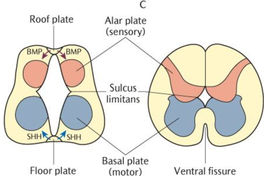
This image shows the cross-section of neural tube. Note the structure of the roof and floor plates compared to the basal and alar plates. From pocketdentistry.com
Development of the spinal cord
Now that we know the structure of the neural tube, the development of the spinal cord is easy.
Sensory structures
The alar plates give rise to the dorsal horns of the spinal cord, which contain sensory neurons whose axons travel to the brain. However, the bodies of the nerve cells of the peripheral nerves lie in the dorsal root ganglia outside the spinal cord. These ganglia arise from neural crest cells and contain pseudounipolar neurons.
Pseudounipolar neurons have one axon which separates into two branches. The central branch grows toward the spinal cord and forms the dorsal (or posterior) root, while the peripheral branch grows toward the periphery. This branch meets the ventral (or anterior) root consisting of axons of motor neurons, forming the spinal nerve. The peripheral axons terminate in sensory receptor organs in the skin, joints, etc.
Motor structures
The basal plates give rise to the ventral horns of the spinal cord, which contain motor neurons. The axons of these neurons form the ventral root, which, after meeting the dorsal root, together form the spinal nerve.
Central canal
The lumen of the neural tube, initially large, becomes smaller and smaller as the mantle and marginal layers grow, eventually reaching the small size of the central canal. The neuroepithelial cells lining the central canal differentiate into ependymal cells.
Summary
- What occurs during neurulation?
- The neural tube and neural crest forms from ectoderm
- Describe the formation of the neural tube
- The notochord induces overlying ectoderm to become the neural plate
- The neural groove forms in the neural plate, causing the lateral edges of the plate to elevate, forming the neural folds
- The neural folds fuse in the midline, initially at the level of the fifth somite
- The neural folds continue to fuse in the cranial and caudal directions
- What are the neuropores, and when do they close?
- After the neural folds have fused, openings remain on the cranial and caudal ends; these are the anterior and posterior neuropores, respectively
- The anterior neuropore closes at day 25, the posterior at day 28
- What are the primary and secondary brain vesicles?
- At the cephalic end of the neural tube three dilations develop; these are the primary brain vesicles, the prosencephalon, mesencephalon, and rhombencephalon
- The primary brain vesicles differentiate into the five secondary brain vesicles, the telencephalon, diencephalon, mesencephalon, metencephalon, and myelencephalon
- What are the three flexures of the neural tube?
- The cephalic flexure, which forms in the region of the mesencephalon
- The pontine flexure, which forms between the metencephalon and myelencephalon
- The cervical flexure, which forms between the myelencephalon and spinal cord
- What is the rhombencephalic isthmus?
- A structure which marks the boundary between the metencephalon and mesencephalon
- What tissue does the wall of the neural tube initially consist of?
- Neuroepithelium
- Describe the structure of the neural tube in cross-section
- An innermost neuroepithelial layer, a middle mantle layer consisting of neuroblasts, and an outermost marginal layer consisting of axons
- The alar plates and basal plates form as thickenings in the mantle layer
- The roof and floor plates are unpaired structures
- What is the function of the roof and floor plates?
- They regulate the organization of the neural tube, especially regulating which neurons should cross the midline
- What gives rise to the dorsal horns of the spinal cord?
- The alar plates
- What gives rise to the dorsal root ganglia of the spinal cord?
- Neural crest cells
- What gives rise to the ventral horns of the spinal cord?
- The basal plates
- What gives rise to the central canal and its ependymal lining?
- The lumen of the neural tube and its neuroepithelial lining