Table of Contents
Page created on August 23, 2020. Last updated on December 18, 2024 at 16:56
Learning objectives
- What are the parts of the brainstem?
- Which brain vesicles give rise to the brainstem?
- Describe the development of the medulla oblongata
- What arises from the basal plates of the myelencephalon?
- What arises from the alar plates of the myelencephalon?
- What arises from the basal plates of the metencephalon?
- What arises from the alar plates of the metencephalon?
- Describe the development of the midbrain/mesencephalon
- What arises from the basal plates of the mesencephalon?
- What arises from the alar plates of the mesencephalon?
- Describe the development of the cerebellum
Development of the brainstem
The brainstem refers to the parts of the brain between the higher centres (cerebrum and cerebellum) and the spinal cord, including the midbrain, pons, and medulla oblongata.
The myelencephalon
The myelencephalon, originating from the rhombencephalon, gives rise to the medulla oblongata. The medulla oblongata, unlike the spinal cord, is not shaped like a tube with the lumen in the middle. Instead, the lateral walls of the tube have moved dorsally, as seen on the image below.
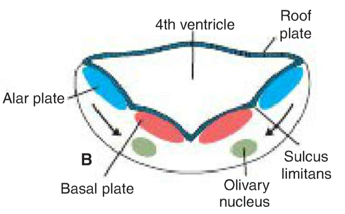
The organization of the medulla oblongata. Note the difference compared to the spinal cord. From Langman’s Medical Embryology.
This forms the 4th ventricle, the CSF-filled space of the medulla oblongata. The dorsal wall of the 4th ventricle consists only of a single layer of ependymal cells, and the pia mater. Together these two layers form the tela choroidea.
The ventral wall of the 4th ventricle is often called the floor of the 4th ventricle.
The basal plates give rise to three types of motor nuclei. From lateral to medial they are:
|
Types of fibres |
Specific nuclei |
| General visceral efferent nuclei |
Dorsal vagal nucleus Inferior salivatory nucleus |
|
Special visceral efferent nuclei |
Nucleus ambiguus |
| Somatic efferent nuclei |
Nucleus of hypoglossal nerve |
The alar plates give rise to three types of sensory nuclei. From lateral to medial they are:
|
Types of fibres |
Specific nuclei |
| Somatic afferent nuclei |
Spinal nucleus of trigeminal nerve |
|
Special visceral afferent nuclei |
Nucleus of solitary tract Vestibular nuclei Cochlear nuclei |
|
General visceral afferent nuclei |
Nucleus of solitary tract |
The metencephalon
The metencephalon, also originating from the rhombencephalon, gives rise to the cerebellum and the pons. Like the myelencephalon and the spinal cord the metencephalon has the basal and alar plates. The alar plates will give rise to the cerebellum, as we’ll see later, but they also give rise to sensory nuclei. The basal plates give rise to the pons and motor nuclei.
The basal plates give rise to three types of motor nuclei. From lateral to medial they are:
|
Types of fibres |
Specific nuclei |
| General visceral efferent nuclei |
Superior salivatory nucleus |
|
Special visceral efferent nuclei |
Motor nucleus of trigeminal nerve Motor nucleus of facial nerve |
|
Somatic efferent nuclei |
Nucleus of abducent nerve |
The alar plates give rise to three types of sensory nuclei. From lateral to medial they are:
|
Types of fibres |
Specific nuclei |
| Somatic afferent nuclei |
Principal nucleus of trigeminal nerve |
|
Special visceral afferent nuclei |
Nucleus of solitary tract |
| General visceral afferent nuclei |
Nucleus of solitary tract |
The name “pons” means bridge, relating to its function of acting as a bridge for nerve fibres connecting the cerebral and cerebellar cortex with the spinal cord.
The mesencephalon
The mesencephalon is the most cranial brain vesicle to retain the basal and alar plates of the neural tube.
The basal plate gives rise to two types of motor nuclei. From lateral to medial they are:
|
Types of fibres |
Specific nuclei |
| General visceral efferent nuclei |
Edinger-Westphal nucleus |
|
Somatic efferent nuclei |
Motor nucleus of oculomotor nerve Motor nucleus of trochlear nerve |
The marginal layer of each basal plate enlarges and forms the crus cerebri, which serve as pathways for nerve fibres from the cerebral cortex to the brainstem.
The alar plates form the anterior and posterior colliculi, which are involved in the visual and auditory reflexes, respectively.
Development of the cerebellum
The dorsolateral parts of the alar plates of the metencephalon bend medially and form “lips”, the so-called rhomboid lips. As the pontine flexure deepens these lips compress, forming the cerebellar plate. The cerebellum develops from this plate. By week 12 the plate has changed shape, now showing a small midline portion (the vermis), and two lateral masses (the lateral lobes)
The cerebellar plate initially consists of neuroepithelial, mantle, and marginal layers, just like the neural tube itself. Some cells of the neuroepithelial layer migrate to the surface of the cerebellum and form the external granular layer. This layer continues to proliferate, giving rise to various cell types of the cerebellum. Some of these cell types migrate back inward.
Purkinje cells originate in the vermis, from which they migrate toward the cerebellar cortex and form the Purkinje cell layer.
Summary
- What are the parts of the brainstem?
- Midbrain, pons, medulla oblongata
- Which brain vesicles give rise to the brainstem?
- Rhombencephalon
- -> metencephalon -> pons, cerebellum
- -> myelencephalon -> medulla oblongata
- Mesencephalon -> midbrain
- Rhombencephalon
- Describe the development of the medulla oblongata
- The lateral walls of the myelencephalon move dorsally, giving the 4th ventricle
- The dorsal wall of the 4th ventricle consists of a single layer of ependyme and the pia mater, together forming the tela choroidea
- What arises from the basal plates of the myelencephalon?
- Motor nuclei of the medulla oblongata, from lateral to medial:
- General visceral efferent nuclei, special visceral efferent nuclei, somatic efferent nuclei
- What arises from the alar plates of the myelencephalon?
- Sensory nuclei of the medulla oblongata, from lateral to medial:
- Somatic afferent nuclei, special visceral afferent nuclei, general visceral afferent nuclei
- What arises from the basal plates of the metencephalon?
- The pons
- Motor nuclei of the pons, from lateral to medial:
- General visceral efferent nuclei, special visceral efferent nuclei, somatic efferent nuclei
- What arises from the alar plates of the metencephalon?
- The cerebellum
- Sensory nuclei of the pons, from lateral to medial:
- Somatic afferent nuclei, special visceral afferent nuclei, general visceral afferent nuclei
- Describe the development of the midbrain/mesencephalon
- The marginal layer of each basal plate enlarges and forms the crus cerebri
- What arises from the basal plates of the mesencephalon?
- Motor nuclei of the midbrain, from lateral to medial:
- General visceral efferent nuclei and somatic efferent nuclei
- What arises from the alar plates of the mesencephalon?
- The anterior colliculi, involved in visual reflexes
- The posterior colliculi, involved in auditory reflexes
- Describe the development of the cerebellum
- Dorsolateral parts of the alar plates of the metencephalon bend and form the rhomboid lips
- The rhomboid lips compress and form the cerebellar plate
- Neuroepithelial cells migrate to the surface of the cerebellum and form the external granular layer
- Cells of the external granular layer give rise to various cell types, some of which migrate back inward
- Purkinje cells originate in the vermis, from which they migrate toward the cerebellar cortex
Hello
In the previous topic you wrote that the metencephalon gives rise to the fourth ventricle, but here you’re saying it comes from the myencephalon.
(Thank you for your notes, saving me right now)
Good catch. I believe it’s both, although the book doesn’t specify explicitly, so I don’t think it’s an important detail anyway. But I’ve fixed the previous topic. Thanks! Good luck!
In the questions it says the myelencephalon gives the motor nuclei of the midbrain but the answer is that it gives the motor nuclei of medulla.. did u mean to write that or is it my misunderstanding ?
Good catch, I think it’s fixed now