Table of Contents
Page created on July 20, 2020. Last updated on December 18, 2024 at 16:56
Learning objectives
- What occurs during gastrulation?
- When does gastrulation occur?
- Describe the process of gastrulation
- Which tissues arise from the ectoderm?
- Which tissues arise from the endoderm?
- What causes the embryonic disc to fold?
- What occurs during craniocaudal folding?
- What is the shape of the embryo after craniocaudal folding?
- What occurs during lateral folding?
- What is the shape of the embryo after lateral folding?
- Which structures form as a consequence of lateral folding?
- What is the notochord and what is its function?
- What is the oropharyngeal membrane?
- What is the cloacal membrane?
- How is the left-right axis of the embryo established?
- Describe the development of the trophoblast during week 3
- Describe the path of oxygen from maternal blood to foetal blood
Gastrulation
Gastrulation is the process where cells of the one-layered epiblast reorganize to form the trilaminar disc, which has three layers. This process occurs during the third week and is the most characteristic event of this week.
The primitive streak
The primitive streak is a narrow and linear band of epiblast cells on the dorsal surface of the epiblast. It appears near the end of week 2 and stretches from the caudal end until approximately half the length of the germ disc. At the cephalic end is the primitive node.
Eventually a narrow groove called the primitive groove develops in the primitive streak. The groove deepens to form the primitive pit.
Invagination
During invagination epiblast cells migrate medially toward the streak and into the primitive pit, forming two new layers of cells beneath the already existing layers of epiblast cells. The bottommost layer is the endoderm, the middle layer is the (intraembryonic) mesoderm, and the layer of epiblast cells which don’t invaginate become the ectoderm.
Each of these layers will give rise specific tissues and organs. These patterns are laid out by the fate map.
Tissues originating from ectoderm
The ectoderm gives rise to the neural tube, the neural crest, the surface ectodermal placodes, and the surface ectoderm, as we will see later. These structures give rise to the organs and structures which maintain contact with the outside world:
- Neural tube
- CNS
- Retina
- Neural crest
- Peripheral nervous system
- Dermis and subcutaneous tissues of the head
- Bones and cartilage of the head
- Odontoblasts
- Melanocytes
- Adrenal medulla
- Surface ectodermal placodes
- Olfactory epithelium
- Inner ear
- Lens
- Surface ectoderm
- Epithelium of the oral cavity, nasal cavity, paranasal sinuses, ear canal
- Epidermis, including hair and nails
Tissues originating from intraembryonic mesoderm
The mesoderm is discussed in topic 3.
Tissues originating from endoderm
The endoderm gives rise to the epithelium of the inside of the body “tube”, as well as the parenchyme of a few organs. More specifically:
- Epithelium of
- GI tract
- Respiratory tract
- Urinary bladder
- Parenchyme of
- Thyroid
- Parathyroids
- Liver
- Pancreas
Folding
At this point in time the embryo is almost disc-shaped and most certainly flat. The embryo will fold along two axes, the craniocaudal axis and the left-right axis.
Both the craniocaudal and lateral folding occur simultaneously, but they’re often discussed separately as it makes them easier to understand. The folding itself occurs because some regions of the embryo grow faster than other, especially the neural tube.
Craniocaudal folding
During craniocaudal folding the embryonic disc flexes along its longitudinal axis, causing it to take on a C shape as the cranial and caudal ends both fold ventrally. Because the yolk sac lies ventrally it will be constricted, causing a portion of it to retract into the embryonic body.
This link contains images and an animation which illustrate the craniocaudal folding well. Click the red arrows in the “Navigation” box to see the state of the folding with each stage.
Lateral folding
During lateral folding the lateral margins of the embryonic disc grow ventrally and fold towards the midline, eventually grasping “around” the craniocaudal axis of the embryo. The lateral margins will eventually converge on the ventral side.
This will give the embryo a more cylindric shape, forming a thick “tube” where the inner lining is endoderm and the outer lining is ectoderm.
After the lateral folding the intraembryonic coelom between the layers of the mesoderm on both sides will communicate. This cavity will give rise to the pericardial, pleural, and peritoneal cavities.
After this folding the gut tube is also formed, which will eventually differentiate into the GI tract. The gut tube maintains an attachment to the yolk sac as the vitelline duct.
Here is the same link for lateral folding.
Notochord
The notochord is a long rod-like structure which is involved in providing molecular signals to the axial skeleton. It induces the formation of the neural plate and later gives rise to the nucleus pulposus of the vertebrae.
In the dorsoventral axis the notochord lies between the endoderm and ectoderm, at the level of the intraembryonic mesoderm. In the left-right axis it lies in the midline of the trilaminar disc.
The notochord arises from epiblast cells which migrate from the primitive node. It eventually spans almost the whole craniocaudal axis of the embryo, but the cranial end is formed first; the caudal end grows afterwards.
The cranial end extends to the prechordal plate, an area just caudal to the oropharyngeal membrane.
The oropharyngeal membrane and the cloacal membrane
The oropharyngeal membrane, also called the buccopharyngeal membrane, is a region at the cranial end of the embryo. It consists of endoderm and ectoderm; at this region there is no mesoderm. This membrane separates the stomodeum, which will give rise to the oral cavity, from the primordial pharynx. Eventually, this membrane will degenerate, which will open the connection between the mouth and the pharynx.
The cloacal membrane is a similar membrane consisting of only endoderm and ectoderm. It lies at the caudal end of the embryo. This membrane separates the upper part of the anal canal (derived from endoderm) from the lower part (derived from ectoderm). Eventually, this membrane will degenerate, which will open the connection between the anal canal and the amniotic cavity.
The left-right axis
After the primitive streak and node has developed it will secrete a signalling molecule called FGF8. This molecule induces expression of a protein called nodal. However, a neurotransmitter called serotonin or 5-HT ensures that nodal is only expressed on the left half of the embryo. On the right half the enzyme MAO degrades serotonin, thereby preventing nodal from being expressed on the right.
Nodal induces another protein called LEFTY2. LEFTY2 activates a transcription factor called PITX2, both of which are only present on the left half. PITX2 is the “master” for establishing the left-right axis.
Abnormalities which cause PITX2 to be expressed on the right half causes defects in laterality, like situs inversus, where the organs usually present on the left are on the right and vice versa, and dextrocardia.
Further development of the trophoblast
When we last checked in on the trophoblast primary villi had just been formed. During week 3 secondary and then definitive villi will form.
A primary villus consists of a cytotrophoblast core covered by a layer of syncytiotrophoblast. During further development mesodermal cells will enter the core of the villus, forming the secondary villus. During even further development these mesodermal cells will differentiate into blood cells and villous capillaries. This is the tertiary or definitive villus.
The villous capillaries will eventually make contact with vessels of the chorionic plate and the connecting stalk. Maternal blood is present in the intervillous space, and nutrients and oxygen from the maternal blood diffuses across the syncytiotrophoblast and cytotrophoblast layers of the definitive villi and into the embryonic blood.
Blood can now travel from the definitive villi and into the embryo. When the heart starts to beat in the fourth week of development, the circulatory system is ready.
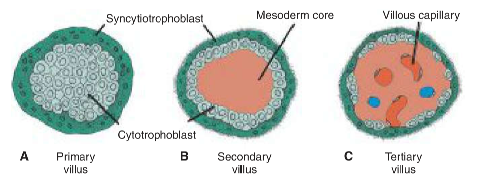
Primary, secondary, and tertiary villi and their structure. From Langman’s Medical Embryology.
Summary
- What occurs during gastrulation?
- Cells of the one-layered epiblast reorganize to form the trilaminar disc
- When does gastrulation occur?
- During week 3
- Describe the process of gastrulation
- The primitive streak and later the primitive groove and pit is formed
- Epiblast cells invaginate into the pit, forming two new layers of cells beneath the already existing layer of epiblast cells
- The most ventral layer becomes the endoderm, the most dorsal layer becomes the ectoderm, and the middle layer becomes the mesoderm
- Which tissues arise from the ectoderm?
- Most components of the CNS and PNS
- Melanocytes
- Adrenal medulla
- Epithelium of oral cavity, nasal cavity, paranasal sinuses, ear canal
- Epidermis, including hair and nails
- Which tissues arise from the endoderm?
- Epithelium of
- GI tract
- Respiratory tract
- Urinary bladder
- Parenchyme of
- Thyroid
- Parathyroids
- Liver
- Pancreas
- Epithelium of
- What causes the embryonic disc to fold?
- Because some regions of the embryo grow faster than other, especially the neural tube
- What occurs during craniocaudal folding?
- The embryonic disc flexes along its longitudinal axis, causing it to take on a C shape as the cranial and caudal ends both fold ventrally
- What is the shape of the embryo after craniocaudal folding?
- It’s a C shape
- What occurs during lateral folding?
- The lateral margins of the embryonic disc grow ventrally and fold towards the midline
- The lateral margins will eventually converge on the ventral side
- What is the shape of the embryo after lateral folding?
- Cylindric, forming a thick “tube” where the inner lining is endoderm and the outer lining is ectoderm
- Which structures form as a consequence of lateral folding?
- The intraembryonic coelom
- The gut tube
- What is the notochord and what is its function?
- It’s a long rod-like structure which provides molecular signals to the axial skeleton
- It induces formation of the neural plate
- What is the oropharyngeal membrane?
- It’s a membrane at the cranial end of the embryo which consists of endoderm and ectoderm
- It separates the primordial oral cavity from the primordial pharynx
- What is the cloacal membrane?
- It’s a membrane at the caudal end of the embryo which consists of endoderm and ectoderm
- It separates the upper part of the anal canal from the lower part
- How is the left-right axis of the embryo established?
- The neurotransmitter serotonin is only present on the left half of the embryo
- On this half serotonin induces nodal, which induces LEFTY2, which induces PITX2
- On the right half of the embryo MAO degrades serotonin
- Describe the development of the trophoblast during week 3
- The primary villi develop into secondary and tertiary villi, the latter of which contain a mesoderm core with capillaries
- These capillaries make contact with the vessels of the chorionic plate
- Describe the path of oxygen from maternal blood to foetal blood
- Oxygen diffuses from maternal blood across the syncytiotrophoblast and cytotrophoblast layers and into the villous capillaries
hi! i just let you know the link that you uploaded here is no longer available i think.
if i clicked it, “page is not found error” like this !
Updated the links now, thanks!