Table of Contents
Page created on July 22, 2020. Not updated since.
Learning objectives
- What are the three subtypes of mesoderm?
- Which tissues arise from the paraxial mesoderm?
- Which tissues arise from the intermediate mesoderm?
- Which tissues arise from the lateral plate mesoderm?
- What is the space between the parietal and visceral mesoderm called?
- What is the septum transversum, and what is its function?
- Which structures separates the pleural and pericardial cavities, and what will these structures give rise to?
- Which structures separate the thoracic and abdominal cavities?
- What are the parts of the diaphragm?
- Describe the formation of the diaphragm
- Why is the diaphragm innervated by a nerve which originates much more cranially?
- From which spinal segments does the phrenic nerve originate?
- Which structures give rise to the serous membranes?
- What are ventral body defects, and what cause them?
- Name some ventral body wall defects
- What are congenital diaphragmatic hernias?
- What is the consequence of congenital diaphragmatic hernias?
Differentiation of the mesoderm
After gastrulation and folding during week 3 the embryo is tube-shaped and consists of three layers – the mesoderm is the middle of these layers.
The mesoderm is divided into three subregions, depending on their position in the trilaminar disc: the paraxial mesoderm, the intermediate mesoderm, and the lateral plate mesoderm.
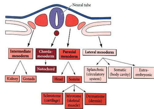
The different mesoderms and their derivatives. Unknown source.
The paraxial mesoderm
The paraxial mesoderm forms clusters of cells called somites which lie on both sides along the craniocaudal axis of the neural tube, hence the name paraxial. There are 35 – 37 pairs of somites, which correspond to the distribution of the spinal nerves. There are approximately 4 occipital somites, 8 cervical somites, 12 thoracic somites, 5 lumbar somites, 5 sacral somites, and 8 coccygeal somites.
Each somite will split into dermatomes, myotomes, and sclerotomes. The sclerotomes will form the bone and cartilage of the vertebrae, the ribs, and the annulus fibrosus of the intervertebral discs. The dermatome will form the dermis and cutis of the back. The myotome will form all skeletal muscles.
The intermediate mesoderm
The intermediate mesoderm gives rise to the genitourinary system, including the kidneys and the gonads. It lies between the lateral plate mesoderm and the paraxial mesoderm, hence the name.
The development of the genitourinary system is described in topics 12, 13, and 14.
The lateral plate mesoderm
The lateral plate mesoderm is the most lateral, hence the name. It splits into parietal (somatic) and visceral (splanchnic) layers. These structures give rise to:
- Parietal mesoderm
- Dermis of the body wall and limbs
- Bones and connective tissue of limbs, sternum, shoulder girdle, pelvic girdle
- Parietal pleura
- Parietal peritoneum
- Parietal pericardium
- Visceral mesoderm
- Cardiovascular system, including heart and blood vessels
- Adrenal cortex
- Spleen
- Intestinal wall, except epithelium
- Visceral pleura
- Visceral peritoneum
- Visceral pericardium
Development of the serous membranes and body cavities
Between the parietal and visceral mesoderm is a space called the intraembryonic coelom, which will differentiate into the body cavities. In the fourth week the intraembryonic coelom divides into the pericardial, pleural, and peritoneal cavities.
The dorsal and ventral mesenteries
After the lateral folding the gut tube will be suspended from the posterior (dorsal) body wall by the dorsal mesentery. It is comprised of visceral mesoderm.
Another mesentery forms, the ventral mesentery. It is also comprised of visceral mesoderm, and it connects the gut tube to the anterior (ventral) body wall. When both the dorsal and ventral mesenteries are present the body cavity is divided into 2 separate halves, but this is only temporary. The ventral mesentery soon disappears, causing the body cavity to be a large continuous space again.
The septum transversum
The septum transversum is a thick plate of mesoderm. It separates the thoracic and abdominal cavities, but not completely, as it leaves the pericardioperitoneal canals on each side of the foregut, the cranial third of the gut tube.
The septum transversum will function as a scaffold for the diaphragm to be formed on.
Separation of the pleural and pericardial cavities and formation of the fibrous pericardium
The pleural and pericardial cavities are separated by the pleuropericardial membranes. These membranes grow out of each side of the lateral body wall. They will eventually fuse, dividing the thoracic cavity into the pericardial cavity and the two pleural cavities. These membranes eventually form the fibrous pericardium.
Separation of the thoracic and abdominal cavities
The thoracic and abdominal cavities still communicate through the pericardioperitoneal canals. These canals will eventually be closed by the pleuroperitoneal membranes, which grow out of the body wall. The membranes eventually fuse, separating the thoracic and abdominal cavities.
Formation of the diaphragm
The adult diaphragm is comprised of three parts: the two crura, the central tendon, and the muscular part. The crura attach the muscular part to the vertebral column. The fibres of the muscular part converge at the central tendon.
The crura (singular: crus) are formed where the pleuroperitoneal membranes fuse with the mesentery of the oesophagus. These membranes continue to grow on top of the septum transversum until they completely cover it and fuse together, forming the central tendon.
The muscular part of the diaphragm originates from myotomes of the cervical somites C3 – C5. The phrenic nerve, which innervates the diaphragm, also originates from cervical segments C3 – C5. During further development the diaphragm migrates caudally, which explains the long course of the phrenic nerve to the diaphragm.
Serous membranes
The serous membranes are the parietal and visceral layers of the three body cavities.
The parietal mesoderm gives rise to the parietal layers of the pleural, pericardial, and peritoneal cavities. The visceral mesoderm gives rise to the visceral layers of these organs.
Relevant malformations
Ventral body wall defects
Ventral body wall defects are developmental malformations where an organ herniates out through the ventral body wall. Most of these are due to the lateral body wall folds not closing properly during the lateral folding. There are several types, depending on the organ affected:
- Ectopia cordis – the heart lies outside the body
- Gastroschisis – intestines lie outside the body
- Bladder exstrophy – the bladder lies outside the body
Omphalocele is another ventral body wall defect involving the intestines, but it is not formed due to improper lateral folding; it’s rather an abnormality of the physiological umbilical herniation of the intestines.
Congenital diaphragmatic hernias
Diaphragmatic hernias involve herniation of abdominal cavity contents into the thoracic cavity. This occurs because the pleuroperitoneal membranes never fully close the pericardioperitoneal canals, leaving a defect in the diaphragm.
Having abdominal contents in the thoracic cavity physically limits development of the lungs, causing the lungs to be impaired at birth. The result is often death.
90% of these hernias occur on the left side.
Summary
- What are the three subtypes of mesoderm?
- Paraxial, intermediate, lateral plate
- Which tissues arise from the paraxial mesoderm?
- Somites
- Dermatomes -> dermis and cutis of back
- Myotomes -> skeletal muscles
- Sclerotomes -> bone and cartilage of vertebrae and ribs
- Somites
- Which tissues arise from the intermediate mesoderm?
- Genitourinary system, including kidneys and gonads
- Which tissues arise from the lateral plate mesoderm?
- Parietal mesoderm
- Dermis of the body wall and limbs
- Bones and connective tissue of limbs, sternum, shoulder girdle, pelvic girdle
- Parietal pleura
- Parietal peritoneum
- Parietal pericardium
- Visceral mesoderm
- Cardiovascular system, including heart and blood vessels
- Adrenal cortex
- Spleen
- Intestinal wall, except epithelium
- Visceral pleura
- Visceral peritoneum
- Visceral pericardium
- Parietal mesoderm
- What is the space between the parietal and visceral mesoderm called?
- Intraembryonic coelom
- What is the septum transversum, and what is its function?
- It’s a thick plate of mesoderm which separates the thoracic and abdominal cavities, but not completely
- It will function as a scaffold for the diaphragm to be formed on
- Which structures separates the pleural and pericardial cavities, and what will these structures give rise to?
- The pleuropericardial membranes
- They will form the fibrous pericardium
- Which structures separate the thoracic and abdominal cavities?
- The pleuroperitoneal membranes, which will close the pericardioperitoneal canals
- What are the parts of the diaphragm?
- Two crura, a central tendon, and a muscular part
- Describe the formation of the diaphragm
- The crura are formed where the pleuroperitoneal membranes fuse with the mesentery of the oesophagus
- The central tendon is formed where the pleuroperitoneal membranes fuse
- The muscular part is formed from myotomes of cervical somites C3 – C5
- Why is the diaphragm innervated by a nerve which originates much more cranially?
- Because the muscular part originates from the cervical somites C3 – C5
- From which spinal segments does the phrenic nerve originate?
- C3 – C5
- Which structures give rise to the serous membranes?
- The parietal mesoderm gives rise to the parietal layers of the pleural, pericardial, and peritoneal cavities
- The visceral mesoderm gives rise to the visceral layers of these organs
- What are ventral body defects, and what cause them?
- They are malformations where an organ herniates out through the ventral body wall
- Most of these are due to the lateral body wall folds not closing properly
- Name some ventral body wall defects
- Ectopia cordis – herniated heart
- Gastroschisis – herniated intestines
- Bladder exstrophy – herniated bladder
- What are congenital diaphragmatic hernias?
- A congenital malformation where abdominal organs herniate through the diaphragm into the thoracic cavity
- What is the consequence of congenital diaphragmatic hernias?
- The lung is compressed, causing abnormal lung development and death