Table of Contents
Page created on October 12, 2020. Not updated since.
Learning objectives
- What is an ion channel?
- What are the different types of ion channels?
- What is membrane potential?
- What are the intracellular and extracellular concentrations of Na+ and K+?
- What is resting membrane potential?
- What is the resting membrane potential of a neuron and a skeletal myocyte?
- What is the function of Na+/K+ ATPase?
- What is the equilibrium potential of Na+ and K+?
- What is an action potential?
- What are the phases of an action potential?
- What causes the cell membrane to depolarize?
- Describe the state of Na+ and K+ channels during the different phases of the action potential
- What is the refractory period, and why is it important?
- What is the myelin sheath, and why is its function in nerve conduction?
- Which cell produces myelin in the CNS and the PNS?
Membrane potential
Ion channels
Ion channels are proteins embedded in the cell membrane that allow ions to be transported across the membrane. Most ion channels only allow one specific ion to pass, most importantly either Na+ or K+, but some ion channels are non-selective, meaning that multiple types of ions can pass through, but only of the same charge.
Some ion channels, the so-called “gated” ones, have “gates” that can be opened or closed, which will allow or deny the ions to pass the membrane. These gates are closed until a specific event occurs, at which point the gate opens, allowing ions to flow through. Many different events can trigger the opening of the gate of the ion channel, depending on the type.
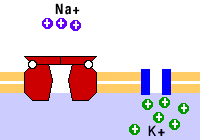
On this figure the red thing is an ion channel that allows Na+ to cross the membrane. This Na+ channel has a gate. The blue thing is a K+ channel. It doesn’t have a gate.
Leakage ion channels have gates which open and close randomly. There is no specific event which opens or closes the gate. Like the name suggests, these “leaky” channels allow ions to “leak” across the cell membrane, even when a specific event has not occurred.
Ligand-gated ion channels have gates which open when a certain molecule binds to the ion channel. The molecule in question is called a ligand, and it is usually a neurotransmitter or a hormone.
Mechanically gated ion channels have gates which open when a physical force is exerted on the cell. This is most characteristically seen in the touch receptors in the skin. When something touches the skin, the cell experiences a force, causing the ion channel to open.
Voltage-gated ion channels have gates which open when the membrane potential of the cell increases (becomes less negative). These are very important for the action potential, as we will see.
Membrane potential
The average cell in the human body has a lot of K+ ion inside the cell, while the extracellular space around it has much less. This is opposite for Na+ – there is way more Na+ in the extracellular space than in the intracellular space. The following table shows the concentration of the two ions in the different spaces in a motor neuron in the spinal cord. The numbers are similar for most cells.
|
Cation |
Extracellular concentration | Intracellular concentration |
| Na+ | 150 mM |
15 mM |
|
K+ |
5,5 mM |
150 mM |
The cell membrane is permeable to K+ in a resting state, but not permeable to Na+. This is because in rest the K+ channels are open while the Na+ channels are closed.
Two forces influence the movement of ions across the cell membrane. The first is diffusion. Diffusion is a force which pushes particles from a solution with higher concentration to a solution with lower concentration. Because the concentration of Na+ is higher outside the cells than inside, there is a concentration gradient across the cell membrane pointing toward the inside of the cell. The opposite is true for K+, so the concentration gradient of potassium points toward the outside of the cell.
The second force is the electrical force. Because there is a different number of charged ions on the inside of a cell and outside, there is also a difference in electric charge between the two. This is called the membrane potential, which once again refers to the difference in electric charge between the inside and the outside of a cell.
In a more physical explanation, it means that the electrical force pushes positive charges (ions) from the outside of the cell to the inside. However, these charges cannot pass freely through the cell membrane, so the force is continuously pushing but there is no (net) flow of positive charges into the cell during rest. Ions can only cross the cell membrane through ion channels.
Resting membrane potential
The membrane potential of a resting cells is aptly called the resting membrane potential. The resting membrane potential of a cell depends on the cell type. It’s -70 mV for neurons, and -90 mV for skeletal and cardiac myocytes. This number means that the inside of the cell is more negative than the outside.
Maintaining the resting membrane potential is very energetically expensive for the cells, consuming up to 70% of the energy requirement of nerve cells! Cells must maintain the high intracellular K+ concentration and low intracellular Na+ concentration by using a protein called the Na+/K+ ATPase, also called the sodium-potassium pump. This protein is in the cell membrane and moves three Na+ ions out of and two K+ ions into the cell with each cycle, each of which consumes one molecule of ATP.
Equilibrium potential
The equilibrium potential is a difficult concept. It is the membrane potential at which the net flow of a specific ion across the cell membrane is zero, because the electric force and the diffusion force are equal and opposite. It’s more important to know the value of the equilibrium potential for each ion than it is to understand the concept:
|
Ion |
Equilibrium potential |
| Na+ |
+60 mV |
|
K+ |
-90 mV |
The resting membrane potential of a cell is very close to the equilibrium potential of K+. This is because at rest the cell is permeable to K+ but not to Na+.
Action potential
An action potential is a series of electrical events in a cell membrane, which causes the membrane potential to quickly increase just above 0 mV and then back to the resting membrane potential. It is what generates nerve impulses in the body. The duration of the action potential is very short, and it depends on the exact cell type. It takes only 1 ms in a nerve cell, but 10 ms in a skeletal myocyte.
During the action potential, some ion channels which were open close and vice versa, creating a net flow of ions across the cell membrane.
Threshold potential
Let’s look at a skin cell which detects touch, a so-called mechanoreceptor. When something touches the skin, mechanically gated Na+ channels open, causing Na+ to flow into the cell. This increases the membrane potential of the cell from -70 mV toward zero, as the difference in electric charge between the inside and the outside of the cell becomes smaller.
When the membrane potential reaches approximately -55 mV, the so-called threshold potential, voltage-gated Na+ channels will open. At this point depolarization occurs.
Depolarization
As soon as the voltage-gated Na+ channels open more Na+ will enter the cell and faster, causing the membrane potential to increase faster. This is called depolarization. The membrane potential continues to increase until it reaches a peak at around +30 mV. During depolarization K+ channels are closed while Na+ channels are open.
The membrane potential peaks because Na+ channels start closing and K+ channels start opening. After the peak, the potential decreases. This is called repolarization.
Repolarization
During repolarization more K+ channels continue to open, while Na+ channels close. The membrane potential decreases as more and more K+ flows out of the cell.
During repolarization, the membrane potential eventually reaches the resting membrane potential (-70 mV), but it doesn’t stop decreasing until it reaches the equilibrium potential of K+ at -90 mV. The membrane potential “undershoots” the resting potential.
Eventually the Na+/K+ ATPase will restore the ion concentration of the resting state, returning the membrane potential to the resting membrane potential.
Refractory period
The refractory period is a period of time in which the cell cannot conduct another action potential at all or with ease. The refractory period is divided into two, the absolute and the relative refractory period.
The absolute refractory period lasts during the depolarization and repolarization. It is absolute because the cell cannot conduct another action potential, no matter the size of the stimulus.
The relative refractory period lasts some time after the repolarization. It is relative because the cell can conduct another action potential during this time, but only if the stimulus is large enough. More specifically, the stimulus must be larger than what’s necessary to cause an action potential in a resting cell. The relative refractory period coincides with the undershooting period, also called afterhyperpolarization.
Myelin sheath
Nerve cells have long “tails” called axons. Most types of nerve cells are myelinated, meaning that their axons are covered in a lipid-rich substance called myelin, forming a myelin sheath. This myelin sheath insulates the axons and significantly increases the speed of which action potentials are conducted along the axon.
The myelin sheath is not continuous; there are gaps in the sheath where the axon is not covered by myelin. These gaps are called nodes of Ranvier.
The myelin sheath of nerves of the central nervous system is produced by a supporting cell type called oligodendrocyte, while the myelin sheath of nerves of the peripheral nervous system is produced by Schwann cells.
Propagation of action potentials in unmyelinated neurons
Action potentials are initiated at one end of the axon. However, the action potential rapidly spreads along the axon, like a wave in the ocean. This is because the Na+ which enters the axon at one end will flow along the inside of the cell membrane, depolarizing that part of the cell membrane, and so on.
Action potentials are only conducted in one direction because the part of the axon which is behind the “wave” is in its refractory period, preventing it from being conducted “backwards”.
Propagation of action potentials in myelinated neurons
Unlike the conduction of action potentials in unmyelinated axons, which spread like a wave, conduction of action potentials in myelinated axons is not continuous but “jumping”. After an action potential arises in one end of the axon, the action potential will “jump” to the next node of Ranvier, where another action potential will arise, and so on.
This occurs because depolarization of one node of Ranvier leads to the opening of Na+ channels in the next node of Ranvier, which depolarized that one, and so on.
TUSEN TAKK!!
🥳
Omg, thanks for all the new topics. When I told you there were some new ones, I thought you meant that you weren’t going to write them because you didn’t like them. So that you wrote them anyways, made me soooo glad! THANK YOU !!!
Glad you like them <3 They're still a work in progress though, many of them are hard to write, and I was never good at muscle physiology