Table of Contents
Page created on October 7, 2019. Last updated on January 24, 2022 at 16:02
Plasma
The plasma is the cell-free part of blood. The plasma accounts for only 55% of the total blood volume, approximately 3000 mL. It’s a pale, yellow, sticky liquid. It’s 90% water and contains:
- Inorganic components
- Ions, like Na+, K+, Cl–, Ca2+, bicarbonate (HCO3–)
- Gases like O2 and CO2
- Organic components
- Proteins
- Albumin
- Globulins
- Fibrinogen
- Lipids
- Carbohydrates
- Glucose
- Amino acids
- Nitrogen compounds
- Urea
- Creatinine
- Uric acid
- Other
- Bilirubin
- Ketone bodies
- Proteins
If you separate the fibrinogen from plasma what remains is called serum. In other words, serum is fibrinogen-free plasma.
The plasma volume can be calculated like this: Plasma volume = blood volume x (1 – haematocrit)
The blood volume can be calculated like this: Blood volume = plasma volume / (1 – haematocrit)
The major plasma proteins
The major plasma proteins are albumin, fibrinogen and globulins. All of them are produced in the liver.
Albumin is a 66 kDa large protein. The normal range is 35 – 48 g/L. It’s a transport molecule. Many compounds bind to albumin in the plasma and are transported with albumin, like Ca2+, indirect (unconjugated) bilirubin, bile salts, fatty acids, lipophilic hormones, medications, etc.
The globulins are a family of globular (globe-shaped) proteins that have higher molecular weight than albumin (>66 kDa). They’re further divided into four categories:
- Alpha-1 globulins – α1-globulins
- Alpha-2 globulins – α2-globulins
- Beta globulins – β-globulins
- Gamma globulins – γ-globulins
The gamma globulins are the immunoglobulins, also known as antibodies. Antibodies are proteins produced by a type of lymphocyte called the plasma cell to fight infections. Antibodies bind to pathogens like viruses and bacteria and make it easier for white blood cells to phagocytose (eat) or destroy them.
There are five subtypes of immunoglobulins, each with different function. You’ll learn more about this in immunology:
- Immunoglobulin type G – IgG – the most abundant immunoglobulin
- Immunoglobulin type M – IgM – the first immunoglobulin to be produced in response to infection
- Immunoglobulin type A – IgA – important to defend against pathogens in mucosa
- Immunoglobulin type E – IgE – important in allergic reactions
- Immunoglobulin type D – IgD
Fibrinogen is a 340 kDa protein. It is essential for blood coagulation. Its function is further described in the blood coagulation topics.
Plasma proteins, especially albumin, are important to maintain the colloid osmotic pressure in the blood. If the concentration of proteins in the blood decreases there is hypoproteinaemia. This can occur in liver disease, kidney disease or if a person doesn’t consume enough proteins. As a consequence, the colloid osmotic pressure in the plasma decreases. This causes water to be “sucked” out of the vessels and into the interstitial space. As a result, interstitial space will swell up. This is called edema.
Serum protein electrophoresis
Electrophoresis is a technique used to separate molecules based on their charge and size. In medicine it’s used to separate the different proteins in the serum, hence serum protein electrophoresis.
It works by putting a sample of serum on a gel. An electric current is applied across the slide. Most proteins have a negative charge and will therefore move slowly through the gel towards the positively charged anode. The smaller and more negatively charged the protein is the further it will travel through the gel.
The result of a normal serum protein electrophoresis looks like this:
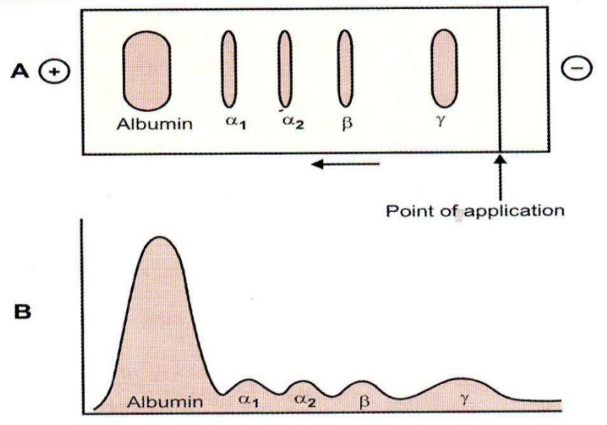 The “point of application” shows where the serum sample was put before the current was turned on. As you can see albumin travelled the farthest, toward the positive anode. The gamma globulins travelled the least and remained close to the point of application. We call each of the “shapes” a fraction. So, there are five fractions.
The size of the fraction on the upper picture, and the size of the peaks on the lower picture both show how much of each fraction there was in the sample. It’s obvious that the most abundant protein in the serum is albumin. The second most abundant are the gamma globulins.
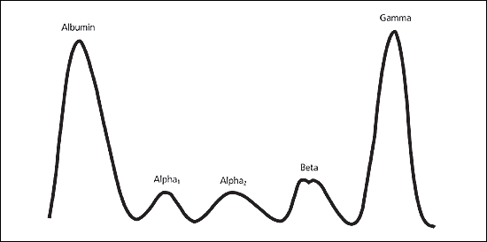
Serum protein electrophoresis in multiple myeloma.
The above picture shows the serum protein electrophoresis of a patient with a disease called multiple myeloma. It is a cancer where cells which produce immunoglobulins proliferate uncontrolledly. These cancer cells produce extreme amounts of immunoglobulins. This is reflected on the picture above, which shows that the serum of this patient contains as many immunoglobulins as albumin.
It’s not important to know about multiple myeloma but it illustrates how serum protein electrophoresis shows the amount of different proteins in the serum.
Acute phase proteins
During an infection or inflammation, the body will elicit a fast, systemic response to combat the infection or tissue damage. This response is called the acute phase reaction. It is this acute phase reaction which produces a fever in response to an infection. You’ll learn more about this in immunology.
During the acute phase reaction certain the production of certain proteins is upregulated or downregulated in the liver. These proteins are called acute phase proteins, simply because their concentration in the plasma changes during the acute phase reaction.
Certain proteins are positive acute phase proteins, because their production is upregulated during the acute phase reaction, so their concentration in the plasma increases. Negative acute phase proteins have their production downregulated, so their concentration in the plasma decreases.
Here are some examples of acute phase proteins:
- Positive acute phase proteins
- C-reactive protein (CRP)
- Fibrinogen
- Negative acute phase proteins
- Albumin
- Transferrin
The fact that the fibrinogen/albumin ratio changes during the acute phase reaction is what explains the erythrocyte sedimentation rate measurement.
Erythrocyte sedimentation rate
If we take a blood sample and just put the tube down in upright position the red blood cells will eventually sink to the bottom of the tube because the iron ions are heavy. The rate at which this sedimentation occurs is called the erythrocyte sedimentation rate (ESR). The rate is normally 2 – 10 mm/hour but the normal value varies with gender and age.
The rate at which this sedimentation occurs depends mainly on the ratio between the protein albumin and the protein fibrinogen in the plasma.
The ESR is mainly used to test for inflammation. During inflammation the acute phase reaction causes the fibrinogen/albumin ratio to increase. This increases the ESR.
Organic non-protein constituents of plasma
The most important organic non-protein constituents of the plasma are shown in the table below:
| Constituent | Example | Function |
| Carbohydrates | Glucose (3.5 – 5.5 mM) | Energy source |
| Other carbohydrates | Energy source | |
| Lipids | Cholesterol | Component of cell membranes, precursor of steroid hormones, precursor of bile acids |
| Triglycerides | Energy source | |
| Free fatty acids | Energy source | |
| Phospholipids | Component of cell membranes | |
| Amino acids | Necessary for protein synthesis, energy source | |
| Urea | Waste product of protein breakdown | |
| Creatinine | Breakdown product of creatine phosphate in muscles | |
| Uric acid | Breakdown product of purine bases | |
| Ketone bodies | Acetoacetate, β-hydroxybutyrate, acetone | Energy source |
| Bilirubin | Breakdown product of haemoglobin |
Viscosity
The viscosity of a fluid is a measure of the fluid’s resistance to flow. When the viscosity is high is the fluid thicker than normal, which means that it is more difficult for the fluid to flow.
The blood is 4-6 more viscous than water, so we say that the relative viscosity of the blood is 4-6. Viscosity is increased when the blood contains more RBCs than normal. This occurs with increased haematocrit and erythrocytosis.
Very viscous blood leads to numerous problems. It elevates the blood pressure, increases the risk for the blood to clot and it can cause headaches.