Table of Contents
Page created on November 7, 2019. Last updated on January 24, 2022 at 16:10
The venous circulation
The venous circulation has two roles:
- To return blood from the tissues to the heart
- To store blood
When we talk about “storing” blood in the venous circulation we don’t mean that venous blood lies stagnated in the venous circulation. We mean rather that the diameter of the veins is very large, which means that the veins contain a lot of blood volume.
Structure of veins:
Veins have a thinner vessel wall than the arteries. Their walls also contain less smooth muscle, but because the pressure in the venous circulation is very low, they can still significantly vasoconstrict.
Veins have valves which prevent blood from flowing backward.
Regulation of venous circulation:
Like the lymph the venous blood is very dependant on skeletal muscle to flow. Venous blood in the lower part of the body must flow against gravity, and because the pressure in the venous circulation is low there is no force which intrinsically pushes venous blood toward the heart. When skeletal muscles around the veins contract the blood is pumped towards the heart.
The veins of the body are innervated by the sympathetic nervous system. When the sympathetic nervous system is activated the veins will vasoconstrict. The blood stored in the veins will be forced back to the heart. The increased venous return will cause an increase in cardiac output thanks to the Frank-Starling mechanism.
This mechanism is important in exercise. In exercise the muscles require more blood, and so the body requires an increased cardiac output. The sympathetic nervous system is activated during exercise, and as we saw above activation of the SNS will increase the cardiac output.
Effect of gravity on the circulation
Blood is pulled down by the circulation. While standing a significant volume of the blood pools in the veins of the legs. When the skeletal muscles are contracting, like when walking, this pooling is reduced.
Valsalva manoeuvre
The Valsalva manoeuvre is when you try to expire against closed glottis/vocal folds. No air will leave the lungs but the pressure inside the thoracic cavity will increase. This is because when you forcible exhale you contract the rib cage, which will compress the air and organs in the thoracic cavity.
The Valsalva manoeuvre is important physiologically in for example defecation and heavy exercise. If performed against a closed mouth and closed nose but with the glottis open air will be forced into the middle ear, which can be relieve ear pain on an airplane.
The Valsalva manoeuvre affects the circulatory system significantly. We can divide the circulatory consequences of the manoeuvre into four phases:
Phase I:
When the intrathoracic pressure increases the large veins, especially inferior vena cava, are compressed by the increased pressure. This decreases the venous return, which will decrease cardiac output by the Frank-Starling mechanism. Both the heart rate and the myocardial contractility decreases.
The aorta is also in the thoracic cavity and will also be compressed during the Valsalva manoeuvre. This causes the pressure in the aorta to increase.
Phase II:
During this phase the aortic pressure will start to decrease due to the reduced cardiac output. The heart rate will increase again.
Phase III:
This phase begins when the patient stops the Valsalva manoeuvre and starts to breathe normally. The aortic pressure will briefly decrease as the intrathoracic pressure is now normalized.
Phase IV:
The venous return returns to normal, so the cardiac output normalizes. The aortic pressure increases, and the heart rate decreases. After some time, the aortic pressure and heart rate will return to normal.
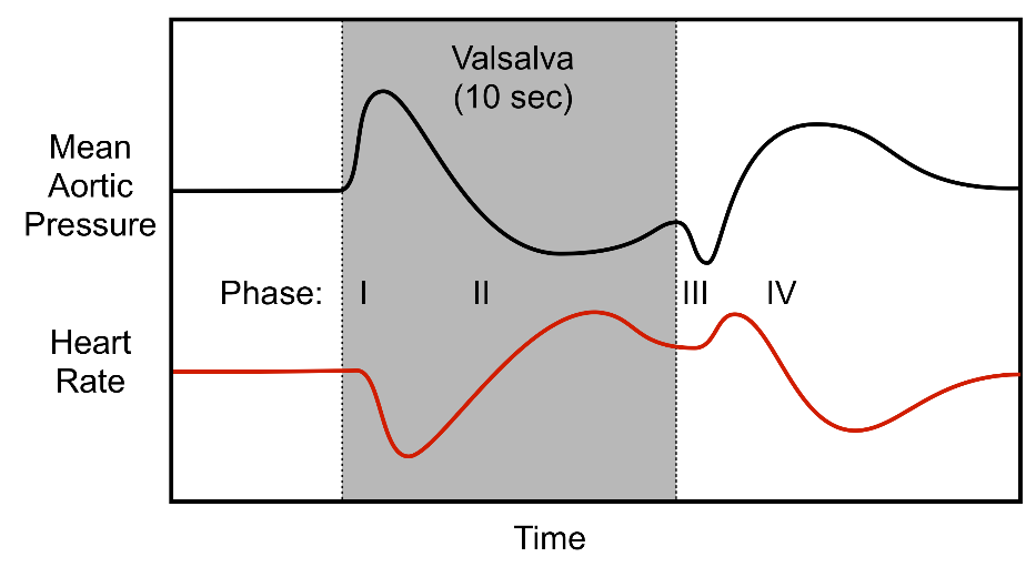
The phases of the Valsalva manoeuvre. The grey area marks where the patient does the Valsalva manoeuvre.
Jugular pulse curve
Some large veins, especially the internal jugular vein, show some pulsation. The pressure inside the internal jugular vein changes with the cardiac cycle, which causes it to pulsate in a specific way. These pulsations can be drawn as a curve.
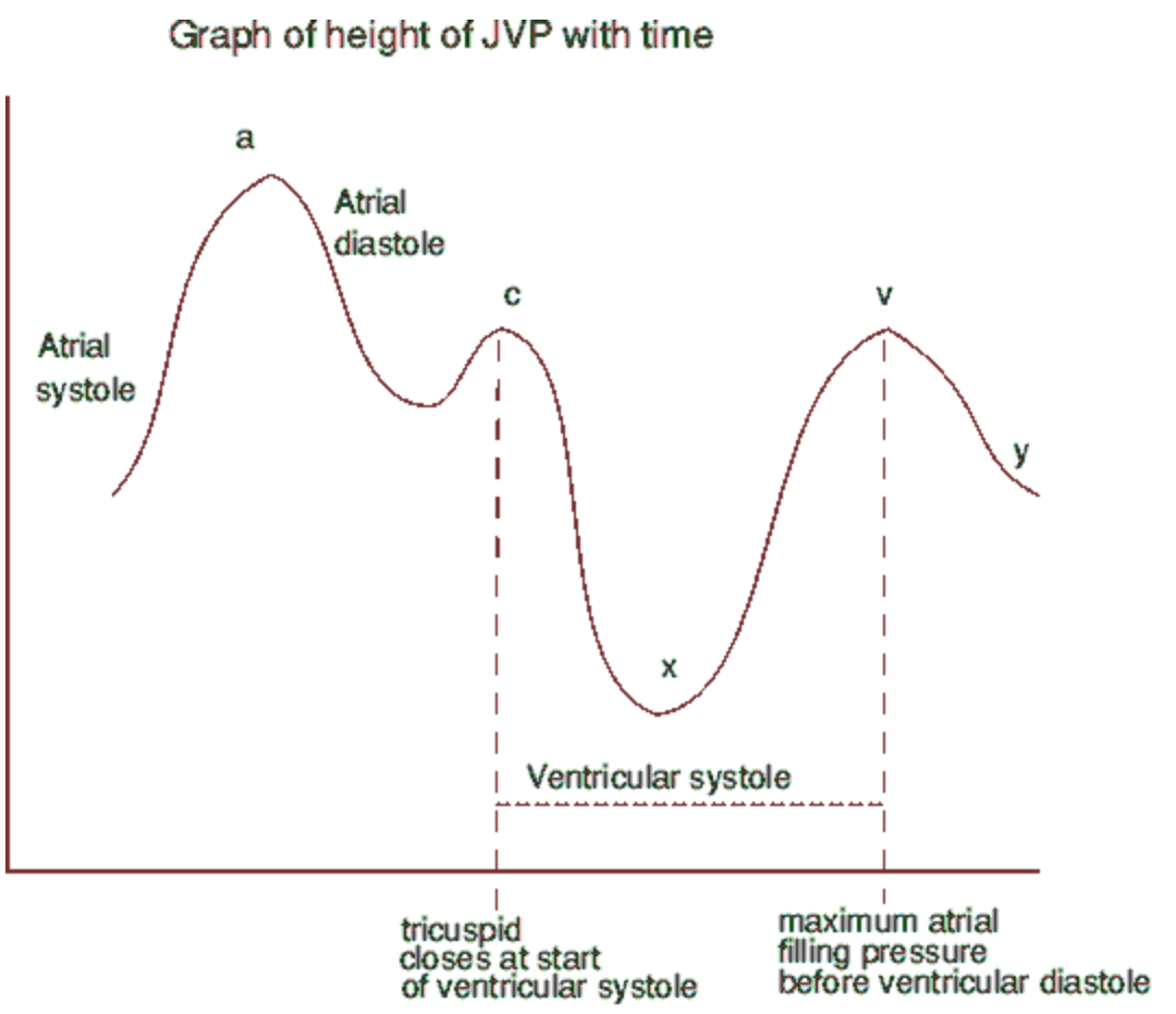
The different waves correspond to different parts of the cardiac cycle:
|
Component of venous pulse curve |
Corresponding event in cardiac cycle |
|
“a” wave |
The end of atrial systole |
|
“c” wave |
The right ventricular contraction, which causes the tricuspid valve to bulge into the right atrium slightly, which causes a small increase in venous pressure |
| “x” descent |
As blood is ejected from the right ventricle, the ventricle takes up less space in the pericardium, causing the right atrium to enlarge |
| “v” wave |
Venous filling of the right atrium while the tricuspid valve is closed |
| “y” descent |
Rapid emptying of the atrium into the ventricle as the tricuspid valve opens |