Table of Contents
Page created on April 19, 2022. Not updated since.
Benign liver tumours and tumour-like lesions
Introduction and epidemiology
The most important benign liver tumours and tumour-like lesions are hepatic haemangioma, hepatic adenoma, and focal nodular hyperplasia.
For pathology, see the corresponding pathology 2 topic.
Clinical features
Benign liver tumours are mostly asymptomatic. The most common symptom is upper right quadrant pain but ruptured or haemorrhaging hepatic adenomas may cause haemodynamic instability.
Diagnosis and evaluation
Asymptomatic liver tumours are usually discovered incidentally on ultrasound or CT imaging.
Hepatic haemangioma is diagnosed in the context of characteristic features on ultrasound, no known neoplastic disease, and normal liver function tests. On CT and MRI with contrast there is a characteristic initial discontinuous enhancement followed by subsequent centripetal enhancement. Haemangiomas must not be biopsied due to the high risk of severe bleeding.
FNH is diagnosed when a homogenous lesion with central stellate scar is visualised on ultrasonography, CT, or MRI. FNH lesions which lack one of the three characteristic classic features on imaging is known as “non-classic” FNH. Non-classic FNH requires biopsy for differential diagnosis.
Unlike hepatic haemangioma and FNH, hepatic adenoma is heterogenous on imaging.
Treatment
Surgical removal of hepatic adenoma is usually advised due to the potential complications, along with stopping contraceptive use. Haemangioma and FNH only require treatment if they’re large, symptomatic, or ruptured.
Cysts of the liver
Introduction and epidemiology
Simple cysts of the liver are usually unproblematic. The clinically important cystic diseases of the liver are polycystic liver disease and echinococcus cyst.
For etiology and pathology, see the corresponding pathology 2 topic.
Clinical features
Liver cysts are mostly asymptomatic. The most common symptom is upper right quadrant pain. Ruptured echinococcus cyst may cause anaphylaxis.
Polycystic liver disease is associated with polycystic kidney disease and therefore kidney failure.
Diagnosis and evaluation
Simple liver cysts are easily diagnosed as thin-walled fluid-filled lesions on imaging. The presence of > 20 liver cysts suggest polycystic liver.
Calcified wall on CT, a positive ELISA, and eosinophilia are characteristic features of echinococcus cyst.
Treatment
Simple asymptomatic liver cysts require no treatment.
Echinococcus cysts should be removed surgically. It’s important to remove the whole cyst without spilling its contents, as it could cause anaphylaxis. Antiparasitic treatment with mebendazole may be considered as the only treatment in small cysts, or as a complement to surgery.
Liver abscess
Introduction and epidemiology
Liver abscesses in Western countries are caused by bacteria, so-called pyogenic liver abscess. In developing countries, liver abscesses are mostly caused by parasites. Liver abscesses mostly affect the right lobe of the liver.
Etiology
In many cases, the source cannot be identified (cryptogenic abscess). In cases where the source is identified, the following sources are the most common:
- Ascending cholangitis (due to biliary obstruction)
- Bacteraemia
- Intraabdominal infection (appendicitis, diverticulitis)
Clinical features
The classical features of pyogenic liver abscess are fever, malaise, and pain in the right upper quadrant. They may also have tender hepatomegaly and may develop jaundice if biliary obstruction occurs. There may also be clinical features of the underlying source.
Diagnosis and evaluation
Elevated inflammatory markers, abnormal liver function test, and positive blood culture is often present.
Imaging shows hypoechoic/hypodense lesions, usually in the right lobe of the liver. An underlying source should also be sought on imaging.
Treatment
Treatment includes antibiotics and drainage, as well as identifying the source. Drainage is often percutaneous, but in complicated cases surgical drainage may be necessary.
Hepatocellular carcinoma
Introduction and epidemiology
For introduction and etiology, see the corresponding pathology 2 topic.
Clinical features
The tumour itself is usually asymptomatic, but the patient usually has symptoms from the underlying disease (cirrhosis or hepatitis). Constitutional cancer symptoms like weight loss, weakness, abdominal pain may be present but may also be caused by the underlying disease.
At later stages, ascites, jaundice, and hepatic failure may develop.
Diagnosis and evaluation
Patients at high risk for HCC (cirrhosis of any cause, chronic hep B) should be screened regularly for HCC. Screening involves measuring abdominal ultrasound and AFP levels. Focal lesions in a cirrhotic liver are primary liver cancer until proven otherwise. AFP is elevated only in 50% of cases, in which case the level correlates with the tumour size.
Contrast CT or MR confirms the diagnosis and shows extrahepatic spread. Typical features on imaging include early contrast uptake, “washout”, and vascular spread.
Liver biopsy is often not needed as the definitive diagnosis can be made with imaging, and biopsy carries a risk of bleeding and tumour spread.
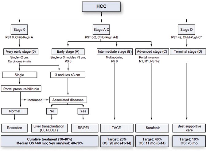
Staging
The Barcelona Clinic Liver Cancer (BCLC) staging is the most frequently used staging system, and at least the one preferred in POTE. The Child-Pugh score and ECOG are important in determining the BCLC stage.
The Child-Pugh score involves multiple parameters that reflect liver function (bilirubin, albumin, PTT, INR, etc.) and classifies the function as Child-Pugh A, B, or C, where A is good liver function and C is poor liver function.
The BCLC staging determines which stage the disease is in, based on the TNM staging, ECOG, and Child-Pugh score:
- Very early stage (stage 0)
- Early stage (stage A)
- Intermediate stage (stage B)
- Advanced stage (stage C)
- Terminal stage (stage D)
Notably, a Child-Pugh C is BCLC terminal stage regardless of the TNM and ECOG features.
The so-called Milan criteria are used to evaluate whether the patient is a candidate for liver transplant or not. The Milan criteria are fulfilled if there is either a single tumour not > 5 cm, or up to 3 tumours, none of which are > 3 cm. There must also be no vascular invasion or extrahepatic spread.
Treatment
There are many treatment options for HCC:
- Resection (preferably laparoscopically)
- Anatomical resection (respecting segmental and lobar anatomy)
- Segmentectomy (or bisegmentectomy or trisegmentectomy)
- Hemihepatectomy
- Sectionectomy
- Atypical resection
- Anatomical resection (respecting segmental and lobar anatomy)
- Liver transplantation
- Local ablation
- Radiofrequency or microwave ablation
- Percutaneous ethanol injection
- Oncotherapy
- TACE (transarterial chemo-embolisation)
- Targeted drug therapy (sorafenib)
The treatment choice depends on the BCLC stage:
From https://abdominalkey.com/liver-transplantation-for-hcc-the-milan-criteria/
Generally, only BCLC stages 0 and A are curable. Curative modalities include surgical resection, liver transplantation, as well as local ablative techniques. Liver transplantation is only considered if the Milan criteria are fulfilled.
Prognosis
Due to the long asymptomatic period, most cases are irresectable at presentation. This gives it a poor prognosis, with a 5-year survival of 30 – 50%.
Other liver malignancies
Intrahepatic cholangiocarcinoma
Intrahepatic CCC is a rare and aggressive liver cancer. It may also develop on the basis of cirrhosis. It spreads along the bile ducts. Treatment is surgical.
Angiosarcoma
Hepatic angiosarcoma is very rare and aggressive. It’s notably associated with exposure to certain toxins, vinyl chloride and thorium dioxide. Vinyl chloride was formerly used in the plastic industry. Thorium dioxide was a radiocontrast material formerly used.
Hepatoblastoma
Hepatoblastoma is a rare childhood tumor that originates in liver precursor tissue, but it’s the most common primary liver tumour in children (0 – 3 years). It usually presents as an abdominal mass.
Secondary liver malignancy
Liver metastasectomy may be used for multiple types of secondary liver cancers, most commonly CRC. Patients with primary colorectal cancer with liver metastasis may undergo metastasectomy with curative intent.
Metastasectomy or debulking may also be used in neuroendocrine, breast, gynaecological, or other secondary liver cancers.
Cholangiocellular carcinoma and gallbladder cancer
Introduction and epidemiology
Cholangiocellular carcinoma (CCC), also called cholangiocarcinoma, is a malignancy of the cholangiocytes that line the bile ducts. It can occur in the extrahepatic or intrahepatic bile ducts, with the former being more common. If a CCC occurs at the confluence of the right and left hepatic bile ducts, it’s known as a Klatskin tumor.
Gallbladder cancer arises from the gallbladder mucosa.
Clinical features
Extrahepatic cholangiocarcinoma presents with signs of cholestasis, including jaundice, pale stool, dark urine, and pruritus. Courvoisier sign (palpable nontender gallbladder) is typical. Intrahepatic CCC and gallbladder cancer is more likely to be asymptomatic and present later.
Diagnosis and evaluation
Tumour markers CA 19-9 and CEA are typically elevated. Ultrasound is good for initial imaging and shows bile duct dilation, but MRCP or ERCP are required for the diagnosis.
Treatment
For cases with resectable disease, radical resection with lymphadenectomy followed by adjuvant chemotherapy is the standard.
Pancreatic cancer
Introduction and epidemiology
Pancreatic adenocarcinoma is a malignancy with one of the highest mortality rates. It’s aggressive, causes no early symptoms, and is difficult to treat. It mostly affects elderly men.
About 85% of cases are irresectable at the time of diagnosis due to being metastatic or locally advanced.
For introduction, etiology, and pathology, see the corresponding pathology 2 topic. For treatment, see surgery topic B55.
Clinical features
Pancreatic cancer generally causes symptoms late, which contributes to the poor prognosis. Tumours in the pancreatic head cause symptoms earlier than tumours in the body or tail.
When symptoms do occur, jaundice, weight loss, anorexia, abdominal pain, vomiting, and constitutional symptoms may occur. Delayed gastric emptying occurs in up to 60% of people with pancreatic cancer and may contribute to the symptoms of anorexia and vomiting. Obstruction of the biliary tract is what leads to jaundice.
Courvoisier sign may be present. It refers to an enlarged, nontender gallbladder with painless jaundice. This contrasts with cholelithiasis, in which the gallbladder is tender.
Trousseau syndrome may also be present. It refers to a migratory, superficial thrombophlebitis.
Diagnosis and evaluation
Abdominal ultrasound is often the first investigation used in patients presenting with jaundice or epigastric pain. US is highly sensitive for pancreatic masses.
Contrast CT or MRI shows the mass and whether it’s resectable or not. It will also show any distant metastases.
The tumour marker CA 19-9 may be elevated; if it is, it may be used for follow-up. Liver enzymes may be elevated if cholestasis has occurred.
The diagnosis requires histological confirmation, which is acquired by biopsy in most cases. However, if the suspicion is high and the mass appears to be resectable, the patient may skip biopsy and proceeds directly to surgery, where the diagnosis is made by intraoperative frozen sections.
My apologies, I can see that you’ve highlighted that (concerning Courvoisier sign) further down!
Not that it’ necessarily so important, I’d just like to point out that Courvoisier sign is a palpable non-tender gallbladder in the presence of jaundice. So having solely a palpable non-tender gallbladder is not a positive Courvoisier sign.
Kind regards 🙂