Table of Contents
Page created on August 17, 2021. Last updated on April 19, 2022 at 10:15
Acute coronary syndrome
Definition
Acute coronary syndrome (ACS) is an umbrella term for acute presentations of coronary artery disease or ischaemic heart disease. ACS is used as an initial working diagnosis until an ECG and troponin level has been performed, which will decide which type of ACS the patient has.
There are three types, each of which with differing underlying pathophysiology:
- Non-ST elevation coronary syndrome (NSTE-ACS)
- Unstable angina (UA)/unstable angina pectoris (UAP)
- Non-ST elevation myocardial infarction (NSTEMI)
- ST elevation myocardial infarction (STEMI)
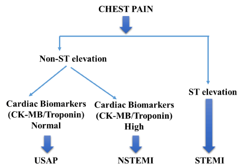
STEMI and NSTEMI are both types of acute myocardial infarction (AMI). This image from here shows the differentiation between the three types of ACS:
Initial evaluation
All patients presenting with sudden onset chest pain or dyspnoea should take an ECG and a blood test to look for troponin levels. STEMI needs acute, urgent management, while NSTE-ACS needs a risk stratification before deciding the timing of management. High risk patients with NSTE-ACS should be treated acutely like STEMI, while low risk patients can be treated on a longer timescale. For this reason, it’s important to determine the type of ACS early, and if it’s either of the NSTE-ACS’s, perform a risk stratification.
The results from the ECG and troponin test will reveal the diagnosis:
- ST-elevations are present in ECG leads corresponding to a part of the heart supplied by a specific vessel -> the diagnosis is STEMI
- ST-elevations are not present in ECG leads corresponding to one part of the heart -> the diagnosis is NSTE-ACS
- Troponins are elevated and are dynamic -> the diagnosis is NSTEMI
- Troponins are not elevated -> the diagnosis is UA
In both STEMI and NSTEMI, the troponin levels increase, peak, and then start to decrease. For this reason, if you measure the troponin level at 2 different times in an AMI, the troponin level is going to be higher or lower at the second measurement, depending on whether the troponin peak had been reached or not at the time of the first measurement. This is called a rise and/or fall in troponin and is often referred to as troponin dynamics. Many conditions can cause troponin elevation, but troponin dynamics are highly specific for AMI. As such, if the diagnosis of AMI is not immediately certain, a repeat troponin measurement should be taken after 1 – 3 hours, and if a rise and/or fall is present, the diagnosis is certain. A significant rise and/or fall is defined as a change of > 20 – 50%.
However, the magnitude of troponin elevation corresponds to the myocardial damage. While conditions like heart failure and myocarditis also can cause troponin elevation, they virtually never cause a very large elevation in troponins. if the troponin levels are very high (even if only measured once), the risk for AMI is sufficiently high that we proceed to invasive management without waiting for a repeat troponin. Modern high-sensitivity troponin assays have an upper normal limit of normal of approx. 14 ng/L. As the single-measurement troponin increases beyond 1000 ng/L the specificity for AMI increases.
Modern high-sensitivity troponin assays can detect a rise in troponins 1 – 3 hours after the infarction. As such, the troponins may be normal if measured early. In these cases, a repeat measurement should be performed 1 – 3 hours later.
![Time course of cardiac troponin elevation as it relates to the size of the myocardial infarction. The time-concentration/activity curves for troponin after large, moderate and small MIs are shown. AMI: Acute myocardial infarction; MI: Myocardial infarction. Adapted with permission from [116].](https://greek.doctor/wp-content/uploads/2021/08/time-course-of-cardiac-troponin-elevation-as-it-re.png)
The change in troponin levels after an AMI over time and depending on size. From here.
An echocardiography should also be performed in the emergency department. Regional hypokinesis or akinesis in regions corresponding to the blood supply territory of one coronary artery increases the likelihood that there is AMI. In addition, echocardiography may reveal alternative diagnoses, like aortic dissection, pericardial effusion, aortic stenosis, hypertrophic cardiomyopathy, or right ventricular dilation suggestive of acute PE.
All patients with suspicion for ACS should have their heart rhythm continuously monitored with a rhythm monitor. The risk for fatal arrhythmias is highest in the first days after an AMI.
ST-elevation myocardial infarction
Definition
An ST-elevation myocardial infarction (STEMI) is defined as the presence of ischaemic symptoms, ST-elevations in a pattern consistent with the blood supply of one coronary artery and a rise and/or fall in troponin levels. Ischaemic symptoms are classically angina, but may also be dyspnoea, nausea, sweating, anxiety, or non-anginal chest pain.
Diagnosis and evaluation
If a patient has typical chest pain and the ECG shows ST-elevations in a pattern consistent with the blood supply of one coronary artery, we do not waste time with a troponin test, as the probability that it is a STEMI is high. However, in many cases a blood test can be performed in the ambulance, thereby not delaying the time to PCI.
These are the ECG leads corresponding to various regions of the heart, and the coronary artery they’re supplied by:
| Leads with ST elevations | Affected myocardium | Supplying coronary artery |
| V1 – V2 | Septal | Proximal LAD |
| V3 – V4 | Anterior | LAD |
| V5 – V6 | Apical | Distal LAD, LCx, or RCA |
| I, aVL | Lateral | LCx |
| II, III, aVF | Inferior | RCA. LCx in 10% of cases |
| V7 – V9 (with reciprocal ST depression in V1 – V3) | Posterior | RCA or LCx |
A new-onset LBBB is considered equivalent to ST-elevation in the case of STEMI. Patients with chest pain and new-onset LBBB are treated as STEMI patients.
ST-depression in leads V1 – V4 may be reciprocal changes of a posterior wall infarct due to a circumflex (CX) artery occlusion. These cases are considered as equivalent to ST-elevation and is treated like STEMI. Posterior ECG leads (V7 – V9) would show ST-elevation.
ST-elevation or depression in the inferior leads (II, III, aVF) suggests inferior wall infarct. In these cases, the right-sided leads V4R, V5R, and V6R should be obtained to assess for a possible right ventricular infarct. In this case, ST-elevation would be present in leads V4R – V6R.
ST-elevations can be present in other conditions which may present similarly, like pericarditis, myocarditis, left ventricular hypertrophy, etc. Care must be used in ambiguous cases, but it’s often better to perform a PCI to be safe.
Pathomechanism
STEMI occurs when a coronary atherosclerotic plaque ruptures, exposing the highly thrombogenic subendothelial matrix of the plaque to the blood. The coagulation system is activated, resulting in a thrombus growing on the ruptured plaque and obstructing the blood flow completely. It is this 100% occlusion which distinguishes STEMI from NSTE-ACS. This occlusion causes a transmural infarction (necrosis) of the myocardium in the area supplied by the occluded vessel.
The process described above refers to the most common type of infarction, the so-called type 1 infarct. Other types of infarcts also exist, most notably type 2 infarcts. In type 2 infarcts there is an increased oxygen demand and/or decreased oxygen supply to the myocardium which is not due to coronary artery thrombosis. This can be due to coronary artery spasm, embolus, tachyarrhythmias, anaemia, respiratory failure, hypertension, hypotension, etc. Up to 28% of patients with myocardial infarction don’t have significant coronary artery disease and so their underlying cause must be something else.
Initial management
If the expected time to a PCI lab is < 120 minutes, the patient is rushed straight there, with as low time from symptom onset to catheter insertion as possible. If the expected time is > 120 minutes, the patient should receive thrombolysis and then be rushed to a PCI lab.
The “classic” acute management for STEMI used to be “MONA”: Morphine, oxygen, nitroglycerine, aspirin. Nowadays we know that morphine may worsen the outcome of patients with AMI, and it should therefore only be given if the patient has strong pain which does not subside after administration of nitro.
Oxygen should only be given if the oxygen saturation is < 90%, as giving it at normal levels of oxygen saturation has not shown any benefit and may even cause harm.
Nitroglycerine, either sublingual tablets or sprays, should only be given if the patient has pain. It may be administered to the patient up to three times but does not improve outcome and is therefore not given routinely to all. Nitroglycerine is contraindicated in right ventricular infarction and should therefore not be administered if right ventricular infarction is suspected.
Aspirin (300 mg) should be administered to all patients to be chewed and swallowed in all cases of STEMI. In addition to this, a loading dose of prasugrel or ticagrelor should be administered.
Beta blocker, most often metoprolol, should be administered in all cases where the patient does not have signs of heart failure and is haemodynamically stable.
Atorvastatin (80 mg) should be administered to all patients as soon as possible.
Unfractionated heparin (UFH) is administered to all patients unless they’re already anticoagulated.
If the anatomy is unsuitable for PCI and the infarcted myocardial area is large, or there is cardiogenic shock, emergency coronary artery bypass surgery (CABG) should be performed.
Summary:
- Aspirin (300 mg loading dose) – in all cases
- P2Y12 inhibitor loading dose (prasugrel or ticagrelor) – in all cases
- Atorvastatin – in all cases
- Unfractionated heparin – in all cases except with pre-existing anticoagulation
- Metoprolol – in all cases except with heart failure
- Nitroglycerine – in case of chest pain
- Morphine – in case of chest pain not responsive to nitro
- Oxygen – in case of O2 sat < 90%
Secondary prevention
See topic 11.
Non-ST elevation coronary syndromes
Definition
A non-ST-elevation myocardial infarction (NSTEMI) is defined as the presence of ischaemic symptoms and a rise and/or fall in troponin levels, but in the absence of ST-elevations.
Unstable angina is defined as the presence of ischaemic symptoms in rest without an elevation of troponin levels beyond the upper normal level.
While ST-elevations are never present in NSTE-ACS (in which case it would be a STEMI), other ECG changes may be present, like ST-depression or T wave inversion. In unstable angina, these changes are transient. ECG changes are a negative prognostic sign.
Pathomechanism
Like STEMI, NSTE-ACS occurs when a coronary atherosclerotic plaque ruptures, exposing the highly thrombogenic subendothelial matrix of the plaque to the blood. The coagulation system is activated, resulting in a thrombus growing on the ruptured plaque and obstructing the blood flow partially. It is this non-complete occlusion which distinguishes NSTE-ACS from STEMI. If this occlusion causes a subendocardial infarction (necrosis) of the myocardium on the area supplied by the occluded vessel, an NSTEMI occurs. If the occlusion is severe enough to cause ischaemia (and resulting symptoms) but not infarction, the diagnosis is unstable angina.
Initial management
The decision of whether to go for an invasive (PCI) or conservative (only drugs) management of NSTE-ACS depends on the patient’s risk stratification. This risk stratification should be done as early as possible. The higher the risk, the sooner they should undergo PCI. Generally, the urgency is evaluated like this:
- Very high risk/unstable patients – PCI within 2 hours
- Drug-refractory angina
- Haemodynamic instability
- Life-threatening arrhythmias
- Mechanical complications
- High risk patients – PCI within 24 hours
- Positive troponin dynamics
- ECG changes
- Low risk patients – Conservative approach initially, PCI electively
- Negative troponin dynamics
- Absence of angina
- Low GRACE-score
Much of the initial management of NSTE-ACS is the same as for STEMI:
- Aspirin (300 mg loading dose) – in all cases
- P2Y12 inhibitor loading dose (ticagrelor only) – in all cases
- Atorvastatin – in all cases
- Unfractionated heparin – in all cases where an invasive approach is taken except with pre-existing anticoagulation
- Metoprolol – in all cases except with heart failure
- Nitroglycerine – in case of chest pain
- Morphine – in case of chest pain not responsive to nitro
- Oxygen – in case of O2 sat < 90%
Secondary prevention
See topic 11.