Page created on November 22, 2018. Last updated on December 18, 2024 at 16:56
More definitions
Metaplasia is the replacement of one healthy cell type to another (but still healthy) cell type which is not usually characteristic for the actual site. This usually occurs in response to some environmental change, and the new cell type is more suited to handle that change. The following examples should be known:
- Barrett metaplasia is a special type of metaplasia in the oesophagus where the normal stratified squamous non-keratinizing epithelium in the lower part of the oesophagus is replaced by simple columnar epithelium with goblet cells, so-called “intestinal” epithelium. This condition is called Barrett’s oesophagus and occurs in response to chronic exposure of the oesophagus to stomach acid, as part of gastroesophageal reflux disease. The intestinal epithelium is better equipped to handle the chronic exposure to gastric acid.
- Squamous metaplasia of the cervical glands
- Metaplasia of the gallbladder in chronic cholecystitis
- Bronchial metaplasia occurs in smokers. The chronic exposure to smoke causes the normal kinociliated pseudostratified columnar epithelium to be replaced by stratified squamous non-keratinizing epithelium
Dysplasia refers to abnormal or disorderly, but not neoplastic, proliferation. It usually occurs in epithelial tissues. The cells don’t change type, but they become abnormal in other ways. Dysplasia usually occurs among the metaplastic cells. The morphology of dysplasia has multiple important characteristics:
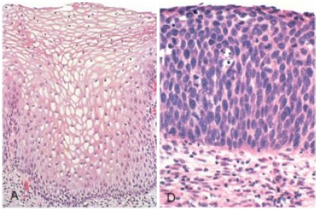
- Loss of maturation and cell orientation is a term used to describe how the cells stop looking and acting like healthy cell. Compare the two pictures of cervical epithelium below. Note how the dysplastic cells don’t look like the healthy cells, they don’t appear to grow in the same way, and they don’t contain the same components (dysplastic cells aren’t filled with glycogen).

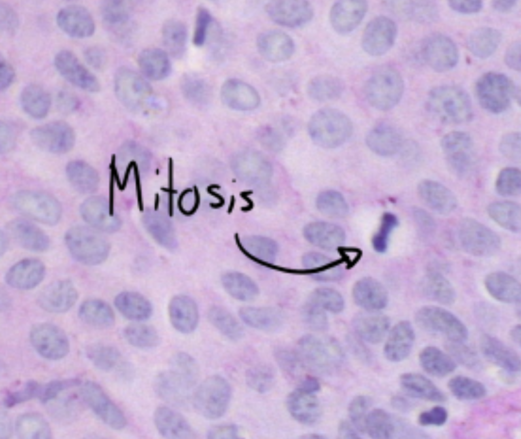
- Mitotic figures are what pathologist call cells that were in the middle of mitosis when the slide was taken. In dysplasia is cell proliferation increased, so mitosis is increased as well.

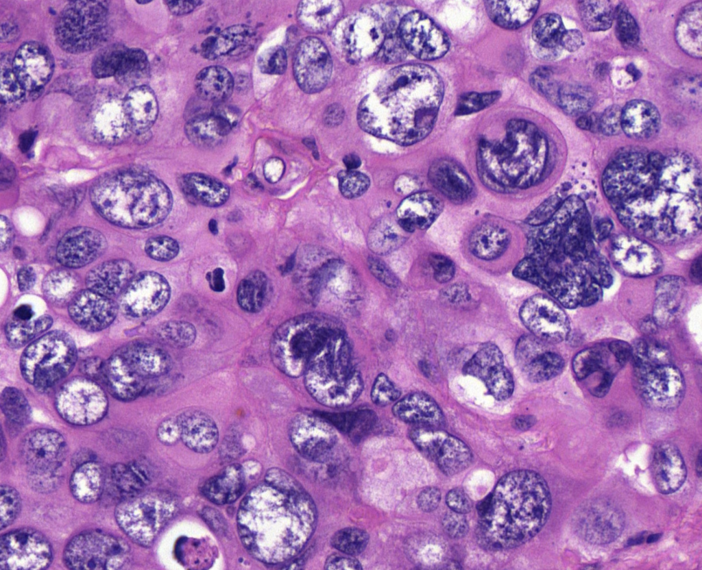
- Pleiomorphism is a term used to describe variability, namely how all the dysplastic cells have variable sizes of cytoplasm (called anisocytosis), sizes of nuclei (anisonucleosis) and how they stain differently (anisochromasia).

- Prominent nucleoli can be present in dysplastic cells. It can be seen in some degree in the “mitotic figures” picture above.
Some important examples of dysplasia:
- CIN
- In Barret’s oesophagus
- Adenomatous polyps in the gastrointestinal tract
Why is metaplasia and dysplasia important? It is because the presence of them often increase the risk for cancer; they are pre-cancerous lesions. Squamous metaplasia in the bronchi due to smoking is an important risk factor for developing lung cancer. Colorectal polyps with dysplasia (adenomatous polyps) have increased risk for developing into cancer, while polyps without dysplasia don’t. The more dysplasia is found in the cervix, the higher the risk for developing cervical cancer.
This warrants an important question; are benign tumors pre-cancerous lesion as well? The answer really depends on which type of benign tumor we’re talking about. The answer is generally no, but it varies from tumor to tumor. Adenomatous polyps are benign tumor-like growths, and they can develop into malignant colorectal cancer. Leiomyomas in the uterus however almost never become malignant.
Hamartoma and choristoma
Hamartomas are benign masses which form from normal but unorganized mature cells. The cells are mature and native to the tissue they grow in, and are therefore not dysplastic nor cancerous. Unlike a true neoplasm, which originates from proliferation of one cell (monoclonal), hamartomas originate from many (polyclonal). A common site of hamartomas is in the lung, where the hamartoma usually contains cartilage, bronchi and fat cells. A hamartoma in the liver could contain hepatocytes, blood vessels or even bile ducts. They are often asymptomatic, however if they grow large enough they could compress nearby structures to cause symptoms. They’re often treated like benign neoplasms.
Choristomas are similar to hamartomas, but that they are organized and not disorganized. The biggest difference is that hamartomas grow in the organs where the cells in the hamartoma belong (like bronchi growing in the lung), but choristomas grow in abnormal locations. An example of a choristoma could be a small nodule of well-developed pancreatic tissue in the submucosa of the stomach, or gastric tissue in the distal ileum.
I don’t get the difference between hamartoma and dysplasia. Help please?
I’ve rewritten the definition, hopefully it’s more understandable now.
Also, leaving your e-mail with the comment causes it to be blocked by the spam filter. I only found your comment by accident.
hy
why leiomyoma cannot be come malignant?
I don’t really know why, but most benign tumors just can’t.
so malignant ones dont related and consequence from benign ones?
Yes, in most cases they’re unrelated.
Burett’s esophagus is listed as an example for both metaplasia and dysplasia; can u please explain that?
Yeah, Barrett’s oesophagus progresses from normal -> metaplasia -> dysplasia -> adenocarcinoma. You can see page 595 in Robbins or here for more info.
Hello there
sry whats the difference btw Neoplasia and Dysplasia?
Dysplasia refers to the abnormal morphology and function of the cell, while neoplasia refers to an uncontrolled growth of cells which will form a tumor. Neoplasia usually occurs with dysplasia and dysplasia is often a precursor for neoplasia.
so only morphology of cells would be the difference?
No, they’re also different in their function and gene expression. To become neoplastic cells must acquire many mutations in genes which usually prevent cells from growing uncontrollably. Dysplastic cells may have some of these mutations, but not enough to allow for uncontrolled growth yet.
Hey,
I am not sure about this but, I think
Hamartomas are abnormally organized cells in normal location and Choristoma are normally organized cells in abnormal locations
Fixed, thanks.