Page created on February 28, 2019. Last updated on December 18, 2024 at 16:56
Staining: HE
Organ: Lymph node
Description:
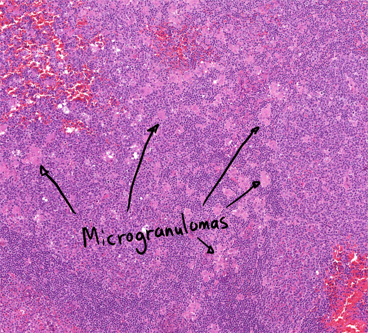
There is extreme(ly large) follicular hyperplasia, as seen by the abnormally large germinal centres. With higher magnification are small granulomas visible, so-called microgranulomas. In a small, triangular area on the right part of the slide can we see B-cells that resemble monocytes, so-called monocytoid B-cell hyperplasia.
Diagnosis: Toxoplasma lymphadenitis
Causes:
- Toxoplasmosis
Theory:
Toxoplasmosis is a condition caused by infection by the parasite toxoplasma gondii. In 90% of cases is the condition asymptomatic. Especially those that are immunocompromised are at risk to develop symptoms, which includes lymphadenitis.
It’s part of TORCH.

Overview
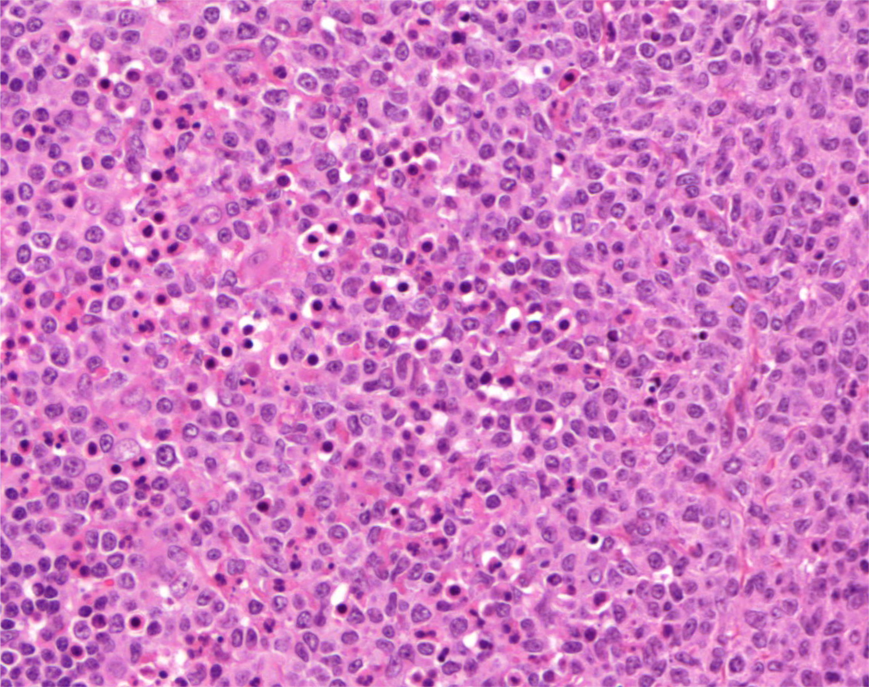
Monocytoid B-cell hyperplasia. From the triangular area on the right part of the slide.