Table of Contents
Page created on September 18, 2020. Not updated since.
Learning objectives
- Describe the formation of the optic vesicle and cup
- What is the choroid fissure?
- Describe the formation of the lens
- What gives rise to the pigmented layer of the retina?
- What gives rise to the neural layer of the retina?
- Describe the formation of the iris
- Describe the formation of the ciliary body
- What gives rise to the choroid?
- What gives rise to the sclera?
- What gives rise to the cornea?
- What gives rise to the vitreous body?
- What gives rise to the optic nerve, and why is it technically not a nerve?
- What does the hyaloid artery give rise to?
Development of visual system
Optic vesicle and cup
At the end of week 4 a pair of outpouchings form on each side of the diencephalon. These are the optic vesicles. As they grow, they come in contact with surface ectoderm, which they will induce to form the lens placodes. Shortly thereafter, the optic vesicle invaginates on itself, forming the optic cup.
The optic cup is double walled. The walls are initially separated by a space, but the space soon disappears, causing the two walls to come into contact with each other. The walls are now known as the inner and outer layers of the optic cup. The optic cup remains connected to the brain by the optic stalk.
During the formation of the optic cup, a groove forms on the inferior aspect. This is the choroid fissure, through which the hyaloid artery enters the eye. The hyaloid artery supplies the lens and retina in intrauterine life before degenerating.
During the seventh week the lips of the choroid fissure fuse, leaving only a round opening at the “mouth” of the optic cup. This opening will give rise to the pupil.
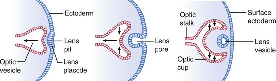
The formation of the lens vesicle and optic cup. The rightmost image shows the situation as the intraretinal space disappears, and the inner and outer layers meet. From https://entokey.com/embryology-pathology/
Lens placode
The lens placode, like all placodes, is a thickening of surface ectoderm. It’s a paired structure which appears in the cephalic region of the embryo by the time neurulation is finished. The lens placode will invaginate and form the lens vesicle. At this point the lens vesicle will lose contact with the surface and instead enter the “mouth” of the optic cup, as seen on the image.
Cells of the posterior wall of the lens vesicle will elongate and gradually fill the lumen of the lens vesicle, forming a structure without a lumen. This gives rise to the lens.
Formation of the retina
The outer layer of the optic cup forms the pigmented layer of the retina, the tenth layer. The inner layer divides into two parts. The posterior four fifths form the pars optica retinae, while the anterior fifth forms the pars ceca retinae.
The pars optica retinae forms the neural layer of the retina, which gives rise to the other nine layers of the retina which aren’t pigmented. This includes the rods and cones.
Formation of the iris and ciliary body
The pars ceca retinae divides into two parts, the pars iridica retinae and the pars ciliaris retinae.
The pars iridica retinae forms the inner layer of the iris. The outer layer of the iris, the pigmented layer, develops from the outer layer of the optic cup. The sphincter and dilator pupillae arise from ectoderm of the optic cup.
The pars ciliaris retinae forms the suspensory ligaments which connect the ciliary muscle to the lens. The ciliary muscle itself forms from mesenchyme which covers the pars ciliaris retinae.
Formation of choroid and sclera
The primordial eye is surrounded by mesenchyme which differentiates into an inner layer and an outer layer. The eye is basically a specialized “elongation” of the brain, so the inner layer of mesenchyme differentiates into a layer comparable with the pia mater of the brain, while the outer layer differentiates into a dura mater-like layer.
The inner layer forms a highly vascularized pigmented layer, forming the choroid. The outer layer forms the sclera, which is continuous with the dura mater of the optic nerve.
Formation of the cornea and anterior chamber
These inner and outer mesenchymal layers form on the anterior aspect of the eye as well but differentiate differently. Between these layers, a lumen forms, which forms the anterior chamber. The mesenchyme anterior to the anterior chamber forms the stroma of the cornea. The posterior mesenchyme forms the iridopupillary membrane, which later disappears.
The adult cornea is formed from this corneal stroma and an inner and outer epithelial layer. The stroma accounts for 90% of the corneal thickness.
Formation of vitreous body
Mesenchyme enters the inside of the optic cup through the choroid fissure. This mesenchyme forms a network of fibres between the lens and retina. The interstitial spaces of this network later fill with a transparent gelatinous substance, forming the vitreous body.
The hyaloid artery, which supplies the foetal lens, goes through the vitreous body. The distal artery of this artery later degenerates, giving rise to the hyaloid canal, which goes through the vitreous body.
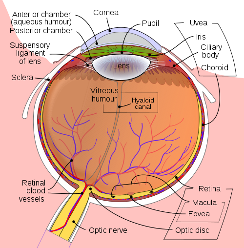
The structure of the eyeball. The hyaloid canal shows where the distal part of the hyaloid artery was. From https://en.wikipedia.org/wiki/Hyaloid_canal#/media/File:Schematic_diagram_of_the_human_eye_en.svg
Formation of optic nerve
As already mentioned, the optic cup is connected to the brain by the optic stalk. The optic stalk gives rise to the optic nerve, which is not technically a nerve but rather an elongation of the brain. This is why the “nerve” is covered by the pia, arachnoid, and dura mater, and why the myelin sheath of the nerve is produced by oligodendrocytes rather than Schwann cells.
Formation of the central retinal artery
The central retinal artery is formed from the proximal part of the hyaloid artery, after the distal part degenerates.
Summary
- Describe the formation of the optic vesicle and cup
- The optic vesicle forms as an outpouching of the diencephalon
- The vesicle invaginates on itself, forming the optic cup
- What is the choroid fissure?
- A groove on the inferior aspect of the optic cup through which mesenchyme and the hyaloid artery enters the eye
- Describe the formation of the lens
- The optic vesicles induce overlying surface ectoderm to thicken and form the lens placode
- The lens placode loses contact with the surface, entering the “mouth” of the optic cup
- Cells of the posterior wall of the vesicle elongate and fill the lumen, forming the lens
- What gives rise to the pigmented layer of the retina?
- The outer layer of the optic cup
- What gives rise to the neural layer of the retina?
- The pars optica retinae of the inner layer of the optic cup
- Describe the formation of the iris
- The iris is formed from the pars iridica retinae of the inner layer of the optic cup, as well as the outer layer of the optic cup
- The pupillary muscles arise from ectoderm of the optic cup
- Describe the formation of the ciliary body
- The suspensory ligaments arise from the pars ciliaris retinae of the inner layer of the optic cup
- The ciliary muscle arises from overlying mesenchyme
- What gives rise to the choroid?
- The inner layer of mesenchyme overlying the eye
- What gives rise to the sclera?
- The outer layer of mesenchyme overlying the eye
- What gives rise to the cornea?
- Mesenchyme anterior to the anterior chamber, as well as epithelium
- What gives rise to the vitreous body?
- Mesenchyme which enters the optic cup through the choroid fissure
- What gives rise to the optic nerve, and why is it technically not a nerve?
- The optic stalk, which is an elongation of the diencephalon and therefore not technically a nerve
- What does the hyaloid artery give rise to?
- The proximal part gives rise to the central retinal artery while the distal part gives rise to the hyaloid canal