Table of Contents
Page created on September 22, 2020. Last updated on May 9, 2022 at 18:18
Learning objectives
- What are the otic placodes, and when do they arise?
- Describe the formation of the otic vesicles (otocysts)
- What are the two parts of the otic vesicles, and which structures will they give rise to?
- Describe the formation of the cochlea and the components inside
- Describe the formation of the semicircular canals
Development of the acoustic and vestibular systems
Otic placodes
The otic placodes appear on each side of the rhombencephalon at day 22 of gestation. They invaginate, first forming the otic pits and then the otic vesicles when they lose contact with the surface. The otic vesicles are also called the otocysts.
Each otic vesicle differentiates into a dorsal, utricular component and a ventral, saccular component. The ventral will give rise to the saccule and cochlear duct. The dorsal will give rise to the utricle, semicircular canals, and endolymphatic duct. The two parts remain connected by the utriculosaccular duct.
Vestibulocochlear ganglion
The vestibulocochlear ganglion originates from cells of the otic vesicles.
Ventral part of otic vesicle: formation of saccule and cochlear duct
In week 6, the cochlear duct starts to grow out from the lower pole of the saccule. It grows in a spiral fashion, until it has completed its infamous 2,5 turns in week eight. While this occurs, in week 7, the cells of the cochlear duct differentiate and form the organ of Corti, which includes the hair cells.
Mesenchyme surrounding the cochlear duct differentiates into cartilage and eventually forms two lumens, the scala vestibuli and scala tympani. These are perilymph-filled spaces, and the scala vestibuli lies above the cochlear duct while the scala tympani lies below it. The vestibular membrane separates the cochlear duct and the scala vestibuli, while the basilar membrane separates the scala tympani from the cochlear duct.
Cells of the saccule differentiate into the sensory cells which sense motions in the vertical plane (movement up and down).
Dorsal part of otic vesicle: formation of utricle and semicircular canals
Also occurring in week 6, the three semicircular canals start to grow out of the utricle as flattened outpocketings. Eventually, the lumen of the outpocketings close everywhere except on the distal end, which will form the canals themselves.
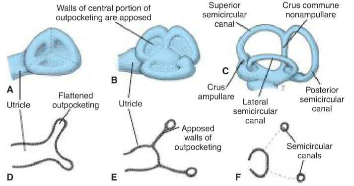
The formation of the semicircular canals. Taken from Langman’s Medical Embryology
Each canal has an ampullary end (crus ampullare) which is dilated compared to the other end (crus nonampullare). In these ampullary ends the cells that sense rotational movement form.
Cells of the utricle differentiate into the sensory cells which sense motions in the horizontal plane (movement forward and backward, and left and right).
Summary
- What are the otic placodes, and when do they arise?
- The otic placodes are thickenings of surface ectoderm which arise at each side of the rhombencephalon
- They arise in day 22
- Describe the formation of the otic placodes (otocysts)
- The otic placodes invaginate, eventually losing contact with the surface, forming the otic vesicles
- What are the two parts of the otic vesicles, and which structures will they give rise to?
- Ventral part -> saccule and cochlear duct
- Dorsal part -> utricle, semicircular canals, and endolymphatic duct.
- Describe the formation of the cochlea and the components inside
- In week 6, cochlear duct grows out from the saccule in a spiral fashion
- Cells of the cochlear duct differentiate and form the organ of Corti
- Mesenchyme surrounding the cochlear duct forms cartilage with two lumens, the scala vestibuli and scala tympani
- Describe the formation of the semicircular canals
- In week 6, semicircular canals grow out of the utricle as flattened outpocketings
- The lumen of the outpocketings will close everywhere except on the distal end, which will form the canals