Page created on October 21, 2019. Last updated on January 24, 2022 at 16:07
The structure of the heart
The heart consists of four chambers, two atria and two ventricles. We can divide the heart into two units, one left unit and one right unit, each unit consisting of one atrium and one ventricle.
Venous blood from the body enters the right atrium through the superior and inferior vena cavae. Blood is pumped from the right atrium into the right ventricle through the tricuspid valve. Blood is then pumped from the right ventricle through the pulmonary valve into the pulmonary circulation. Inside the pulmonary circulation CO2 will be released from the blood while the blood will become saturated with O2.
Oxygenated blood from the pulmonary circulation enters the left atrium through the pulmonary veins. Blood is pumped from the left atrium into the left ventricle through the mitral valve. From the left ventricle blood is pumped through the aortic valve into the aorta, where oxygen-rich blood will reach all parts of the body.
We call the valves between the atria and ventricles the atrioventricular valves or AV valves. This includes the mitral and the tricuspid valve. We call the valves between the right ventricle and pulmonary trunk and between the left ventricle and the aorta the semilunar valves. This includes the aortic and pulmonary valve.
The cardiac cycle
The cardiac cycle is the sequence of events that occurs during 1 heartbeat. Its length depends on the heart rate. If the heart rate is 60 bpm the cardiac cycle lasts for one second.
The cardiac cycle consists of two parts: the systole and the diastole.
The diastole is the part of the cardiac cycle where the heart muscle is relaxing and is filling up with blood. It normally accounts for 2/3 of the duration of the cardiac cycle in rest, but when the heart rate increases the diastole becomes shorter (but the systole stays more or less the same).
The systole is the part of the cardiac cycle where the ventricular muscle contracts. The duration of the systole is not significantly changed when the heart rate changes.
We can divide the cardiac cycle into 7 phases.
Phase 1 – isovolumetric contraction:
Phase 1 marks the beginning of the systole. At the beginning of this phase the ventricles are filled with blood. All four heart valves are closed. During this phase the ventricles contract, but because the atrioventricular and semilunar valves are closed, the blood can’t go anywhere. Instead, the pressure inside the ventricles increases due to the contraction.
The name of this phase comes from how the volume of the ventricles doesn’t change.
Phase 2 – fast ejection:
When the pressure inside the ventricles becomes high enough the semilunar valves will be pushed open, allowing fast ejection of the blood into the pulmonary trunk and the ascending aorta. This occurs when the pressure inside the left ventricle exceeds 80 mmHg and the pressure inside the right ventricle exceeds 8 mmHg. The aortic valve opens because the pressure inside the left ventricle becomes higher than the pressure inside the aorta. The pulmonary valve opens because the pressure inside the right ventricle becomes higher than the pressure inside the pulmonary trunk.
About 70% of all blood which is ejected by the ventricles is ejected during this phase. The remaining 30% of the blood is ejected during the next phase.
Phase 3 – slow ejection:
Blood continues to be ejected by the ventricles but at a slower rate than in the fast ejection, because the pressure inside the ventricles begins to decrease. The aortic pressure also decreases because the blood which is pumped out is now leaving the large arteries and reaching the small arteries. The slow ejection lasts twice as long as the fast ejection.
This phase marks the end of the systole.
Phase 4 – isovolumetric ventricular relaxation:
This phase marks the beginning of the diastole. The ventricles suddenly stop contracting and start relaxing, which causes the ventricular pressure to decrease rapidly. The semilunar valves close because the pressure inside the aorta and pulmonary trunk is now larger than the pressure inside the ventricles. The volume of the ventricles doesn’t change during this phase, hence the name.
Phase 5 – rapid ventricular filling:
The pressure inside the ventricles has now decreased so much that the ventricular pressure is less than the atrial pressure. This causes the atrioventricular valves to open. The large pressure difference between the atria and the ventricles cause blood to flow from the atria to the ventricles.
It is during this phase when most blood enters the ventricles from the atria.
Phase 6 – slow ventricular filling (diastasis):
Phase 6 is the longest phase of the cardiac cycle. Blood continues to flow from the atria to the ventricles, but because the pressure difference between the two is now smaller this filling is much slower than in the previous phase. Only a small amount of blood enters the ventricles during this phase.
When the heart rate increases, this phase is the phase which is shortened. This makes sense because during this phase not much blood flows from the atria to the ventricles anyway, so shortening this phase doesn’t reduce ventricular filling significantly.
Phase 7 – atrial systole:
During this phase the atria contract, pumping blood through the atrioventricular valves into the ventricles. This contributes to, but is not essential for, ventricular filling of blood. The atrial systole increases the ventricular volume by only 5%. In fact, the heart can function normally without atrial systole because this phase contributes so little to the ventricular filling.
The semilunar valves are closed. This is the last phase of the diastole.
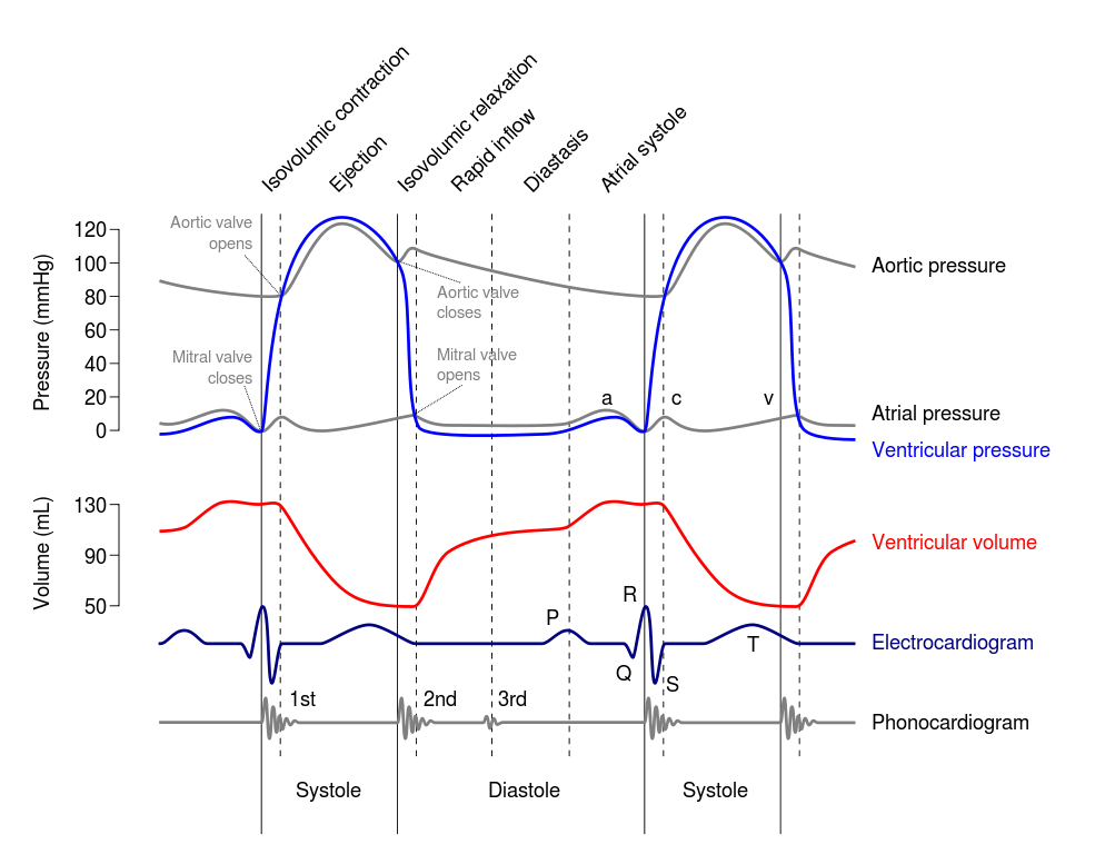
This is called a Wiggers diagram. It shows how the different parameters of the left atrium, left ventricle and aorta changes during the different phases of the cardiac cycle.
Events of the cardiac cycle:
|
Phase |
Phase name | Part of the cardiac cycle | Semilunar valves | Atrioventricular valves | Corresponding EKG wave | Corresponding heart sound |
|
1 |
Isovolumetric contraction | Systole | Closed | Closed | QRS | S1 |
|
2 |
Fast ejection | Systole | Open | Closed | – | – |
|
3 |
Slow ejection | Systole | Open | Closed | T | – |
| 4 | Isovolumetric ventricular relaxation | Diastole | Closed | Closed | – |
S2 |
| 5 | Rapid ventricular filling | Diastole | Closed | Open | – |
S3 (pathological) |
| 6 | Slow ventricular filling | Diastole | Closed | Open | – |
– |
| 7 | Atrial systole | Diastole | Closed | Open | P |
S4 (pathological) |