Table of Contents
Page created on October 19, 2020. Not updated since.
Learning objectives
- What is a synapse?
- Describe how an electrical synapse works
- Describe how a chemical synapse works
- What is the presynaptic and postsynaptic membrane?
- What is a neurotransmitter?
- What is the synaptic cleft?
- Which are the most important neurotransmitters, where in the body are they found, and which receptors do they activate?
- How is acetylcholine removed from the synaptic cleft?
- What are neuropeptides?
- What are the types of cell membrane receptors? Explain their mechanisms
- What is EPSP and IPSP?
- Which are the excitatory neurotransmitters?
- Which are the inhibitory neurotransmitters?
- What is spatial summation and temporal summation?
This is quite an important topic for understanding physiology well, so I recommend you spend time to understand it. Otherwise, you should be good if you know the learning objectives.
Neurochemistry of synapses, neurotransmitters, postsynaptic receptors, and neuromodulators
So far, we’ve looked at how nerve signals are transmitted across axons, but that’s only a piece of the puzzle. Most electrical signals originate or terminate in the brain, but no one axon goes from the brain and to a muscle or any other organ. Multiple nerve cells are connected in series, one after another. We’ll now look at how communication between nerve cells occurs.
Synapses
A synapse is an area where action potentials are transmitted from one nerve to another nerve or to a muscle. Synapses can be between the axons, dendrites, and the neuron soma, or any combination of them. An axodendritic synapse is between the axon of one neuron and the dendrite of another. An axoaxonic synapse is between the axons of two neurons. An axosomatic synapse is between the axon of one neuron and the soma of another.
According to the mechanism of transmission, there are two types of synapses: electrical synapses and chemical synapses.
Electrical synapses
Electrical synapses are simple. Gap junctions connect the nerve cell to the next, allowing ions to pass through from the first cell to the second. This transmits the action potential between cells in the simplest way.
Electrical synapses are less important than chemical synapses for us medical students and doctors.
Chemical synapse
Chemical synapses are more complex and unfortunately more important to know about. In a chemical synapse, when the action potential has reached the “end” of the axon or dendrite of the first nerve cell, the cell membrane at the “end” will be depolarized. This depolarization triggers the release of a certain molecule, which will be released into a small space between the two nerve cells. The molecule will then diffuse to the next cell, where it is recognized by its specific receptor. This will initiate a reaction there, most commonly the generation of a new action potential, which conducts the signal along. The neurotransmitter is then broken down or taken up by one of the cells.
I use these terms in singular form, but in reality many, many neurotransmitters are released and bind to many, many receptors.
There are some new terms to be known here. The part of the cell membrane of the first neuron which is involved is called the presynaptic membrane, while the part of the cell membrane of the second neuron involved is called the postsynaptic membrane. The space between these membranes is the synaptic cleft, which is approximately 20 nm wide. The molecule which is released is called a neurotransmitter. We say that the chemical synapse consists of the presynaptic membrane, the synaptic cleft, and the postsynaptic membrane.
The neuromuscular junction is another name for the chemical synapse between the axon of an alpha motoneuron and a skeletal muscle fibre. This is the most important type of chemical synapse for us to know, but it’s explained in the next topic.
There are many different neurotransmitters, each of which have separate functions, roles, and effects. For each neurotransmitter there is a set of receptors which are in the postsynaptic membrane which recognizes them. Let’s look at the most important ones.
Acetylcholine
Acetylcholine is probably the most important neurotransmitter. Chemical synapses with acetylcholine are found in the brain, in the neuromuscular junction, in preganglionic and postganglionic parasympathetic synapses, and in preganglionic sympathetic synapses.
There are two receptors which are activated by acetylcholine. They are the muscarinic acetylcholine receptor and the nicotinic acetylcholine receptor. The former is such named because it’s also activated by a poison produced by the toadstool mushroom called muscarine, while the second gets its name from the fact that it’s also activated by nicotine. There are multiple subtypes of muscarinic acetylcholine receptors, but we won’t get into that now.
After the signal has been transduced to the postsynaptic neuron and acetylcholine has done its job, it will be degraded in the synaptic cleft by an enzyme called acetylcholinesterase.
Norepinephrine
Norepinephrine, also called noradrenaline, is both a hormone and a neurotransmitter. Chemical synapses with norepinephrine are found in the brain and in postganglionic sympathetic synapses.
There are two receptors which are activated by norepinephrine. They are the α-adrenergic and β-adrenergic receptors. There exist multiple subtypes of each of these receptors.
Dopamine
Dopamine is also both a hormone and a neurotransmitter. Chemical synapses with dopamine are mostly found in the brain.
Dopamine activates the simply named dopamine receptors, of which there are several subtypes.
Other less important neurotransmitters
Aspartate, GABA, glutamate, glycine, serotonin, endorphins, and nitric oxide are all neurotransmitters which are important, but they’re considerably less important for us now. Nitric oxide is perhaps the most interesting of these as it is the only one which is a gas rather than a solid. You’ll learn more about nitric oxide as a neurotransmitter in biochemistry.
Neuropeptides
Neuropeptides are small peptides which are involved in the synapses. In most cases they’re released together with neurotransmitters and either increase or decrease their effect, both in the short-term and in the long-term. In some cases, neuropeptides act as neurotransmitters themselves.
Some important neuropeptides include VIP, neuropeptide Y, enkephalin, somatostatin, cholecystokinin, etc. Many neuropeptides are also hormones in the gastrointestinal tract.
The effect of a neurotransmitter on the postsynaptic membrane
As already mentioned, when the neurotransmitter is recognized by its specific receptor on the postsynaptic membrane the receptor will be activated. This causes a reaction in the postsynaptic neuron. The specific reaction depends on the type of neurotransmitter and receptor.
There are two possible consequences of a chemical synapse. Either the postsynaptic membrane becomes more positive or more negative. This is called EPSP and IPSP, respectively. But first, we should discuss a bit about receptors.
Cell membrane receptors
Cell membrane receptors are proteins which are embedded in the cell membrane, spanning its whole thickness, which certain molecules can bind to and activate. It has two important parts: the part on the extracellular side and the part on the intracellular side. A molecule which is recognized by a receptor is called a ligand.
The extracellular part of the receptor is what recognizes the ligand. After a ligand has bound to this part, the conformation of the whole receptor changes. This triggers an intracellular response, which the intracellular part of the receptor is responsible for.
There are two types of receptor: ionotropic receptors and metabotropic receptors.
Ionotropic receptors are also called ligand-gated ion channels, because they are both receptors and ion channels. These ion channels are normally closed, but open when a ligand binds to them. The nicotinic acetylcholine receptors are ionotropic receptors, as well as some of the receptors for GABA, glycine, and glutamate.
Metabotropic receptors are more complex. When the ligand binds to the metabotropic receptor the intracellular part of the receptor does something which causes an intracellular response. The most common subtype of metabotropic receptor is the G-protein coupled receptors. When activated, these receptors will activate a so-called G-protein. This G-protein will then go on to trigger one or more mechanisms inside the cell, some of which may cause opening of ion channels. Metabotropic receptors are much slower than ionotropic receptors.
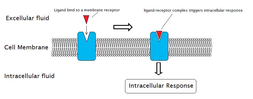
The general mechanism of a cell membrane receptor. From Wikipedia.
You’ll learn more about these receptors in biochemistry.
Excitatory postsynaptic potential
Excitatory postsynaptic potential (EPSP) refers to when the neurotransmitter causes the postsynaptic membrane to be excited. This means that the membrane potential of the postsynaptic membrane will increase, inching closer to the threshold value for depolarization. An EPSP occurs if the activation of the receptor on the postsynaptic membrane causes an influx of positive ions (typically Na+) in the postsynaptic membrane.
Neurons are connected to each other in very complicated networks, meaning that one neuron will synapse with many other neurons. So-called multipolar neurons have many dendrites, each of which the axon of another neuron can synapse on. Let’s imagine a neuron which has 3 chemical axodendritic synapses to its dendrites.
Let’s say that each of these synapses can increase the membrane potential of the neuron by 5 mV. Recall that the resting membrane potential of a neuron is -70 mV, and the threshold for depolarization is -55 mV.
If only two of the axodendritic synapses are activated, the postsynaptic membrane potential will increase by 10 mV, from -70 to -60 mV. This is not enough to cause depolarization, but the membrane is now so hypopolarized that it only needs 5 mV more to depolarize. If the last of the synapses is also activated, the postsynaptic membrane will reach threshold, causing it to depolarize.
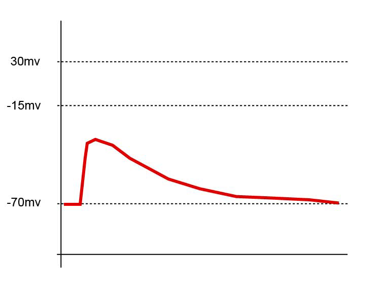
This figure shows the first scenario, where the membrane is hypopolarized but not enough to cause a depolarization and formation of an action potential. On this figure the threshold is -15 mV. From Wikipedia.
That’s not to say that more than one chemical synapse is always necessary to depolarize the postsynaptic membrane: one can be enough. It depends on the specific synapse and neurons. However, you can think of an EPSP as increasing the tendency for the postsynaptic membrane to produce an action potential.
There are certain neurotransmitters which are always excitatory, meaning that they always produce an EPSP (rather than an IPSP). They are glutamate and aspartate. Acetylcholine is excitatory if the acetylcholine receptors on the postsynaptic membrane are of the nicotinic type. If the postsynaptic membrane contains muscarinic acetylcholine receptors, the result can be either excitatory or inhibitory.
IPSP
Inhibitory postsynaptic potential (IPSP) refers to when the neurotransmitter causes the postsynaptic membrane to be inhibited. It’s the exact opposite of the EPSP, decreasing the tendency for the postsynaptic membrane to produce an action potential.
An IPSP occurs when the neurotransmitter causes negative ions to flow into the postsynaptic membrane, or positive ions to flow out of it. This hyperpolarizes the membrane, reducing the membrane potential to lower than the resting membrane potential. This makes it harder for the postsynaptic membrane to reach the threshold value.
GABA and glycine are inhibitory neurotransmitters, meaning that they always produce an IPSP.
Signal integration and summation
A nerve cell of the central nervous system, for example an alpha motoneuron in the spinal cord, has connections with hundreds or thousands of other neurons through synapses. We can think of these as signal inputs. The process of determining whether to fire an action potential or not based on the hundreds or thousands of excitatory and inhibitory inputs is known as signal integration.
EPSPs and IPSPs are not permanent changes in the postsynaptic membrane; they last only for a short amount of time. For two inputs (EPSPs or IPSPs) to influence each other, they must either occur simultaneously or in rapid succession. This is called summation, of which there are two types: spatial summation and temporal summation.
Spatial summation occurs when two inputs arrive simultaneously. The resulting membrane potential will be the numerical sum of the two inputs. If one of the inputs is an EPSP which increases the membrane potential 10 mV, and the other input is an IPSP which decreases it with 5 mV, the sum is an increase in in membrane potential of approx. 5 mV. Likewise, EPSPs and IPSPs of equal magnitude can cancel each other out. Spatial summation requires inputs from multiple neurons.
Temporal summation occurs when two inputs arrive not simultaneously, but right after each other. Despite not arriving at the same time these inputs can be summed, as long as the second input before the first input disappears. Unlike spatial summation, temporal summation can occur with only one input. This is because the postsynaptic potential lasts longer than the interval between action potentials. The former lasts approx. 15 ms, while the refractory period of an action potential is only approx. 1 ms.
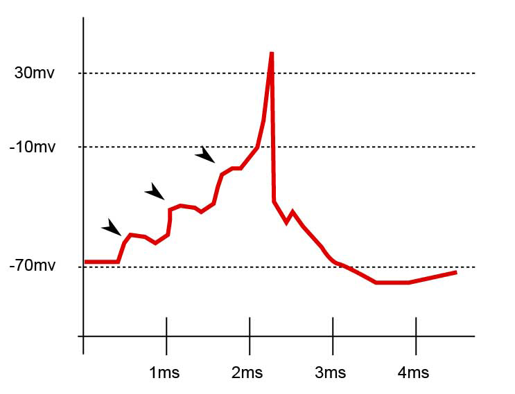
This figure shows the temporal summation of three EPSPs, which together causes the membrane potential to hit the threshold, causing depolarization and generation of an action potential. On this figure the threshold is – 10 mV. Each arrow shows the time when one EPSP arrived. From the same source as above.