Table of Contents
Page created on October 14, 2018. Last updated on October 14, 2021 at 15:52
The composition of this topic is random.
Hypoplasia
Incomplete development or underdevelopment of an organ or tissue.
Aplasia
Aplasia refers to defective development of an organ or tissue beyond an early embryonic stage. As a result, the embryonic remnants of the organ are often present.
Agenesia
Agenesis refers to complete loss of an organ or part due to development never beginning in the embryonic stage.
Atrophy
Atrophy means that an organ or a tissue has been reduced in size due to a decrease in cell size and number. There are many causes of atrophy, and it may seem like too much to remember, but if you think over it, they are pretty logical. Let’s take a look at them.
- Decreased workload – Atrophy of disuse
- This happens for example when you are immobilized for some time or if you used to work out a lot, and then stop. Your muscles become weaker and smaller because you didn’t use them. These situations are reversible since the cells decreases in size. However, if this immobility is prolonged, the cells will undergo apoptosis and there will be a decrease in both cell number and size.
- Loss of innervation – Denervation. Neuropathy
- This is loss of nerve supply that will result in muscle atrophy.
- This can happen if you experience some trauma or an inflammation.
- Diabetes and alcohol can also affect the innervation.
- Some causes are hereditary or rare tumors of nerves.
- Diminished blood supply – Ischemia
- Slowly developing arterial occlusions like atherosclerosis is the main cause for ischemia.
- Cells in the brain and the kidneys are especially affected for this type of atrophy.
- This is also called a senile atrophy as it happens in older people.
- Inadequate nutrition
- This is caused by severe protein- and calorie malnutrition, also called marasmus, and usually occurs in children in developing countries.
- The cells begin to use muscle as energy source because there is no fat left, leading to muscle atrophy.
- The end result will be cachexia.
- Loss of endocrine stimulation
- During menopause a physiological atrophy will happen in the endometrium, vaginal epithelium and breast due to less stimulating hormones. They are no longer needed, and the tissue will undergo apoptosis.
- Taking exogenous steroids can make the cortex of adrenal gland regress because it’s no longer used.
- Pressure atrophy
- Ischemic changes due to an enlarging benign tumor that will compress the arteries.
- For example: an aneurysm can cause pressure on vertebrae, giving atrophy of the vertebrae.
- Other example: deposition of amyloids causes pressure atrophy of cells
Mechanisms of atrophy
In atrophy, the cell size and the organelles inside the cells are decreased. The cells must reach a new equilibrium at a lower level. The cells will also have limited function, but they are still not dead. However, if there is an ischemia the process may reach a point of irreversible change and go through apoptosis. How does this atrophy happen? Let’s look at the pathomechanism.
When a cell is not used or doesn’t get enough nutrition, the protein synthesis and the metabolic activity of the cell will decrease to make its size smaller, because an unused cell is a waste of energy to maintain. The cell will also increase the protein degradation by using the ubiquitin-proteasome pathway. Intracellular proteins will get tagged with ubiquitin and destroyed by proteasomes.
The cell will also use autophagy to decrease in size. Cellular components will be packed in in vacuoles, and then fuse with lysosomes for digestion. Some of the cell debris is resistant to digestion, like lipofuscin, leading to a brown color as seen in brown atrophy of the heart. This is regulated by the autophagy genes.
Gross morphology and microscopic characteristics
Brain atrophy
The causes for the brain tissue to regress is atherosclerosis or typical degenerative brain disorders as Alzheimer’s disease.
In atrophy of brain it’s possible to see that the sulci are wider, and the gyri are narrower. The ventricles will also look bigger. Because of smaller brain mass, the amount of CSF will increase to compensate the empty space, leading to hydrocephalus ex vacuo. You can see brain atrophy here.
Brown atrophy of the heart

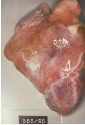
This is typically caused by cachexia, where the nutrition is not enough, and the body will begin to use muscles as fuel. The muscle mass of the heart will decrease, making the coronaries twisted. The subpericardial fat is also drastically decreased.
The heart will get a teardrop-like shape, be smaller and look brown because of lipofuscin accumulation.
Kidney atrophy
Usually caused by narrowing of the renal artery, due to atherosclerosis. The kidney will shrink because of ischemia. The decreased blood flow will also activate the renin-angiotensin-aldosterone system, because the kidney will think the blood pressure is low in the whole body. This will lead to hypertension. The other kidney must compensate and will go through hypertrophy. You can read more about this process here.
Osteoporosis
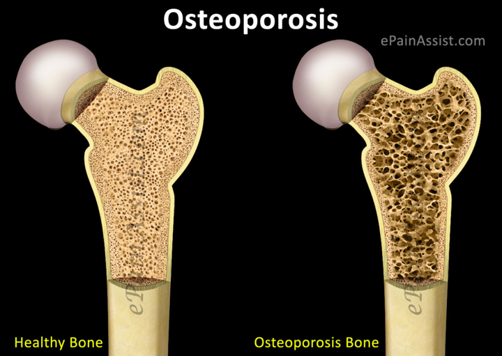
Osteoporosis is a boring disease making your bones very fragile and bone fractures will happen easily, especially in the area of the neck of femur. This fragility comes from a decrease in bone mass.
Its age-related, and progress more and more each year. The most common forms are senile and postmenopausal. Let’s take a look at them:
Senile
Osteoblast activity will decrease with age, and with decreased physical activity and decreased bone formation, the bones will get very fragile.
Postmenopausal
Estrogen makes sure that the osteoclasts are inhibited by inducing apoptosis for them. However, after the menopause the estrogen levels drops, and there’s suddenly not enough estrogen to keep the osteoclasts away from the bones. This increased bone resorption leads to fragile bones and osteoporosis, so tell your grandma to be careful!
Is there a difference in loss of organ between aplasia and agenesia?
Changed the wording slightly to hopefully make the distinction clearer.